Two legs in the same patient — and the blinded assessor who found no difference
A randomized trial treated one leg with Nd:YAG laser and the other with polydocanol foam in the same patient. The dermatologist who performed the two gave different notes to both legs; blind evaluators, looking at the same photographs, found no difference (P 0.84).
 View the video lesson
Telangiectasias of the lower limbs are extremely common: a German epidemiological study found telangiectasis or reticular veins isolated in 59% of the general adult population. In more than half of the cases, symptoms related to venous hypertension appear throughout evolution — it is not only an aesthetic issue.

The intrapatient design
Fifty-six women with primary telangiectasis and reticular veins (C1A), phototypes I to III, between 23 and 66 years old (mean 46) were included. Patients with varicose veins above C1, occlusive arterial disease or pregnancy were excluded, and all underwent duplex — none had reflux greater than one second in deep veins, saphenous junctions, trunks, taxos, or perforating. If the veins were not symmetrical between the legs, the patient was excluded. One leg was drawn for Nd:YAG laser with long synchronized pulse; the other received foam sclerotherapy. Fifty-one of the 56 completed the study.
The laser used fluence from 100 to 200 J/cm2, spot from 3 to 7 mm and pulse width from 10 to 50 ms, with cooling by cold air; the technical outcome was clinically observable closure of the vessel during irradiation. Sclerotherapy used 0.5% polydocanol in foam, 2 mL syringe and 30G needle — with transillumination, also reaching the feeding reticular. Two sessions with 6 weeks interval, 26 to 32 mmHg compression medium during the day for 3 weeks, and reassessment at 6 weeks and 6 months.
The finding that matters: who judges the result
The clearance was scored on a six-point scale (1 = no improvement; 3 = 30-40%; 4 = 50-70%; 5 = above 70%; 6 = 100% bleached). In the final evaluation of the dermatologist who performed both treatments, the laser leg was median 3 (IQR 2) and the foam leg was median 4 (IQR 2), significant difference (P) < 0.001). The two blind evaluators, who did not participate in the clinical phase, scored the same photographs with median 5 (IQR 1) for both legs.
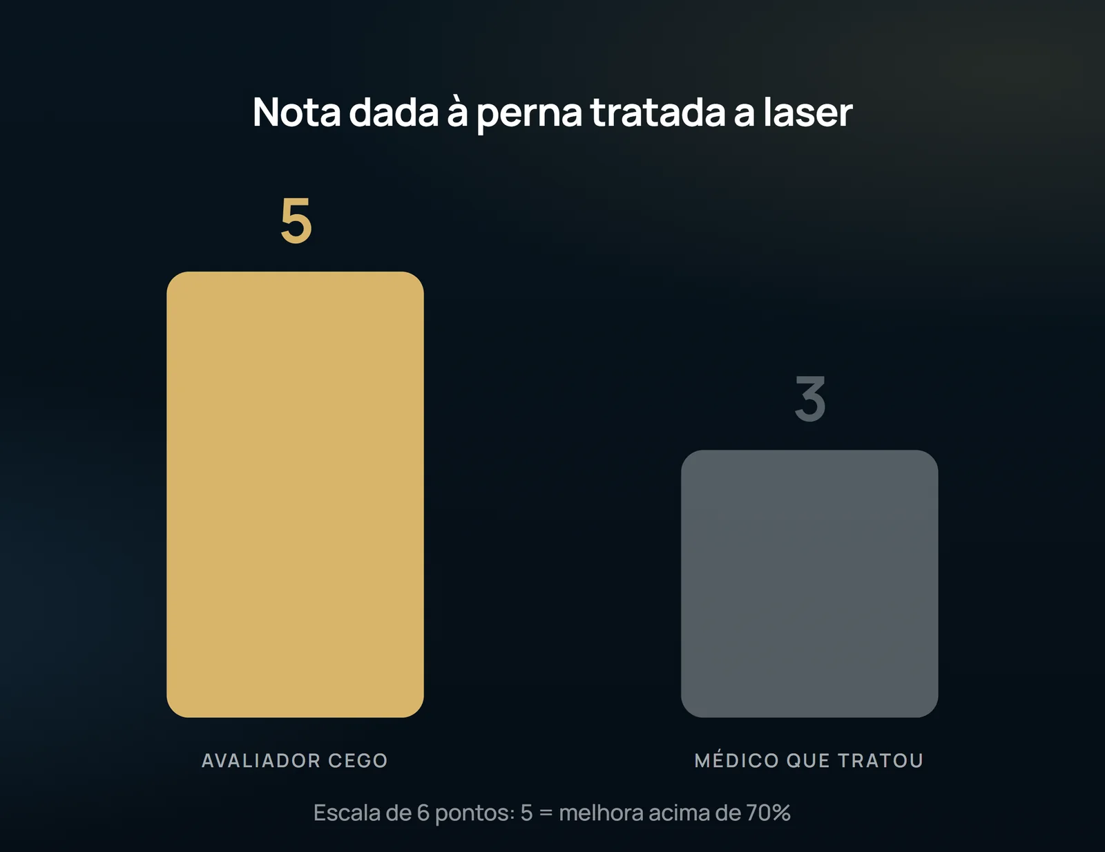
The authors themselves explain the discrepancy: the non-blind dermatologist had many years of experience with sclerotherapy and felt more confident with it. Sclerotherapy was, in fact, faster to clear — both for the doctor who treated and for the blind — but by the last visit, the difference had disappeared.
Side effects: then the score reverses
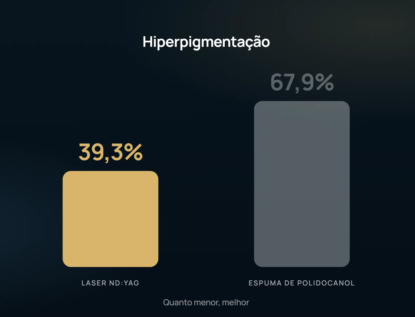
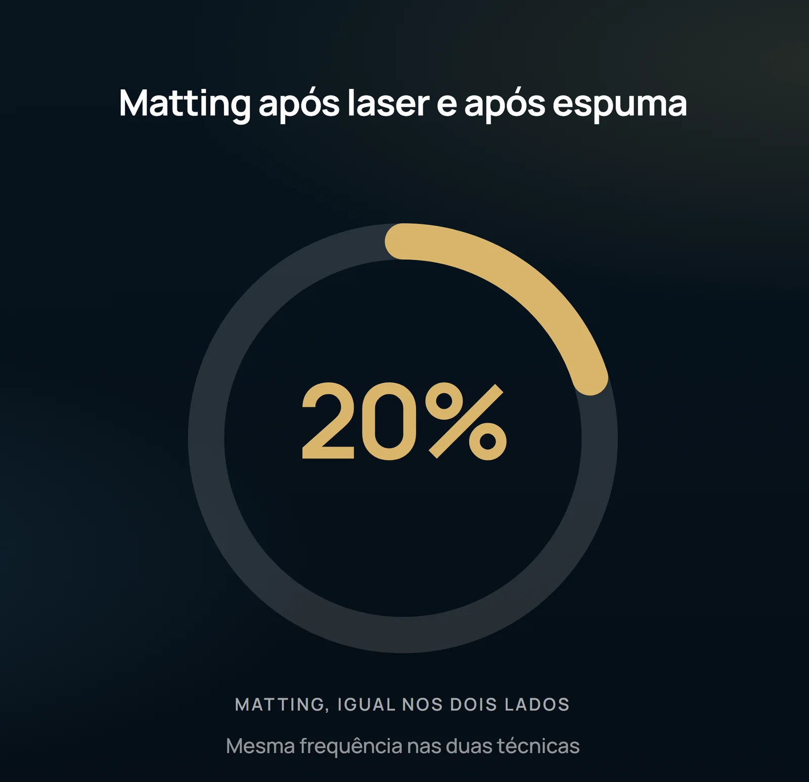
The authors attribute the high rate to the choice of foam: the foam displaces the blood, comes into full contact with the endothelium and adheres better to the wall, being more effective than the liquid form — and for the same reason, associated with more pigmentation. Matting occurred with equal frequency in both techniques, in 20%. Limited cutaneous necrosis appeared in one patient in each group. Satisfaction was equally high on both sides (median 5, IQR 2; P 0.58 in subjective perception of improvement). There was a significant difference in pain: the laser was felt as more painful (P 0.003).
What the study does not authorize to state
The trial is open — neither patients nor the performing physician were blind, and it was precisely this absence of blinding that produced the central finding. The population is narrow: only women, phototypes I to III, C1A, without stem reflux, with telangiectasis of 0.2 to 1 mm and reticulars of 1 to 2.9 mm, and symmetry between the legs as a requirement for inclusion. None of this is transported to insufficient saphenous skin, to more pigmented skin or to men. The follow-up is 6 months after the second session: there is no long-term recurrence, and hyperpigmentation, although usually regressed within 6 to 24 months, may persist more than one year. The outcome is photographic and ordinal, not an objective measure of occlusion. And the result "no difference" is no difference detected with 56 patients — no proof of equivalence.
Content addressed exclusively to health professionals. No individual result is guaranteed: the indication depends on clinical evaluation and imaging examination of each patient (CFM 2,336/2023).