Laser in one leg, glucose in the other: when photographs and patients disagree
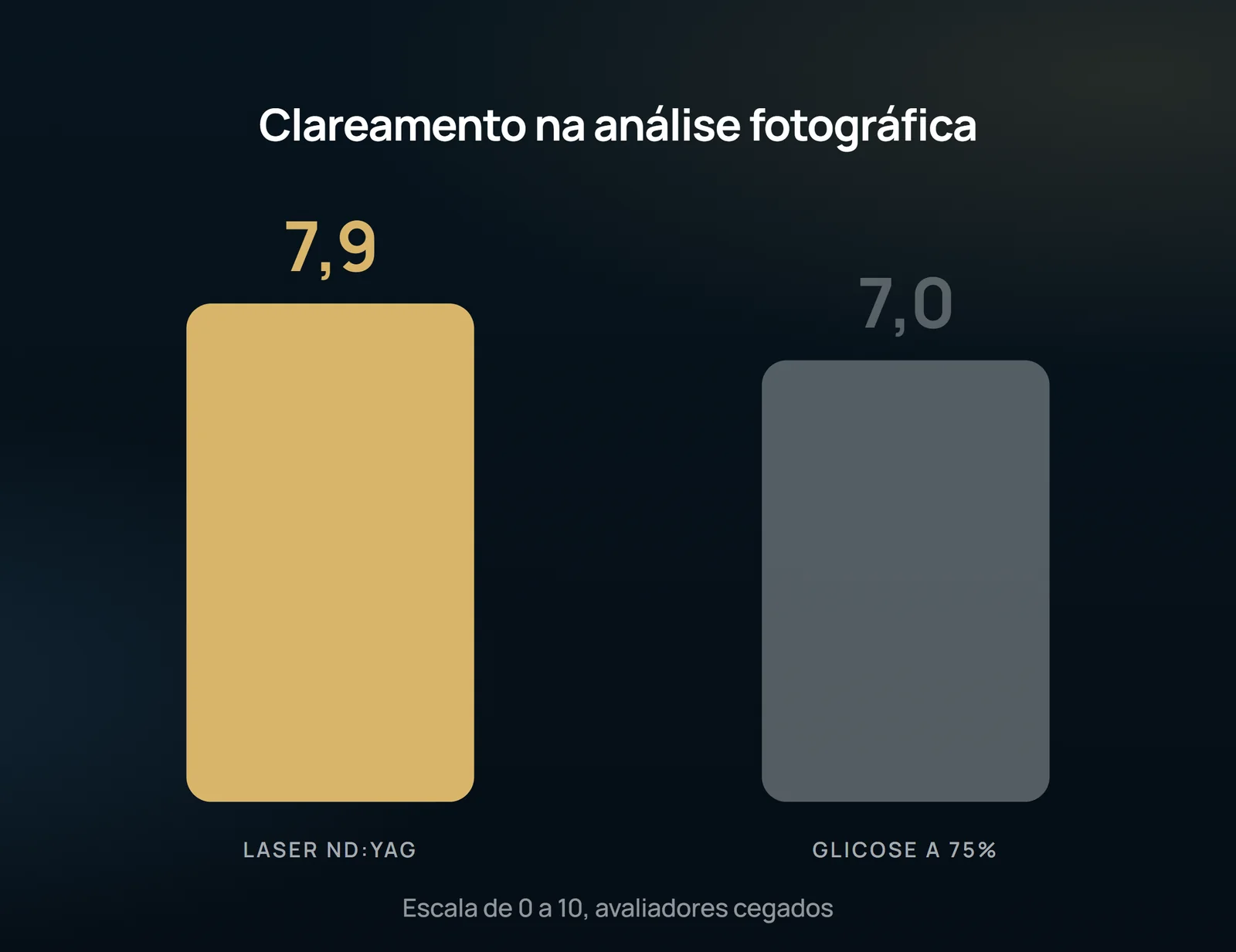
Thirty women, one leg drawn for each method, three sessions and two blind evaluators. In the photographic analysis, the laser clarified more (7.9 × 7,0). In pain and satisfaction, sclerotherapy has won. The article concludes that both treat equally well.
 View the video lesson
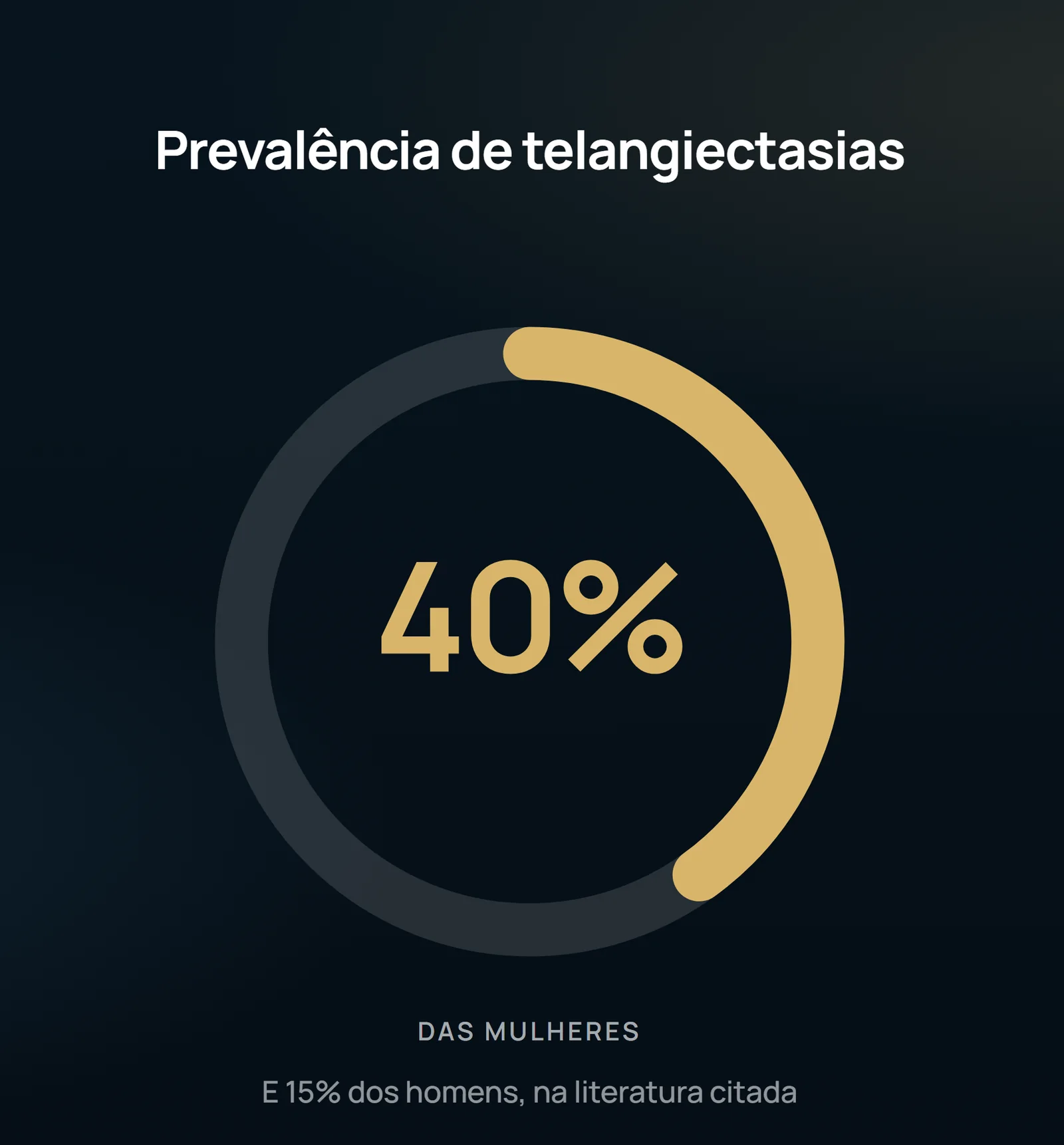
Lower limb telangiectasia affects approximately 40% of women and 15% of men. Sclerotherapy, used since 1930, remains the gold standard treatment for low cost and good results, usually with hypertonic glucose 75% per needle 27 or 30 G. The laser entered the dispute after Anderson and Parrish described selective photothermolysis in 1983 — but few studies compared the two prospectively, and none had compared Nd:YAG with hypertonic glucose in the same patient over three sessions.
The intrapatient design
Thirty women aged 25 to 65 years (mean 43), with phototypes I to VI and telangiectasis from 0.5 to 1.5 mm in diameter, were included. All patients were CEAP C1: patients with evident signs of chronic venous insufficiency or varicose veins (C2 and above), pregnant, lactating or anticoagulation were excluded. In each patient, one leg was randomly assigned to laser (Xeo, Cutera) and the other to sclerotherapy with 75% glucose solution at comparable ankle, calf, popliteal fossa and thigh sites. The same doctor applied the two modalities in three sessions with an interval of one month.

The laser was applied with a cooled spot of 3 mm to 1 Hz, fluence of 100 to 120 J/cm2 and pulse width of 15 ms (smaller vessels) or 30 ms (larger vessels), with skin cooling before and after pulses; the final points were darkening of the target vessel and perivascular erythema. Sclerotherapy was performed with low flow injections, 3 mL syringe and 30 G needle, until complete clearance of the vessel, totaling 1 to 2 mL per session, without posterior compression and with guidance to avoid sun for 7 days. The photographs were taken before and 1 week after treatment and scored from 0 (no improvement) to 10 (complete clarification) by the applicator physician and two blinded independent observers.
The objective outcome favors laser
Reported outcomes favor glucose
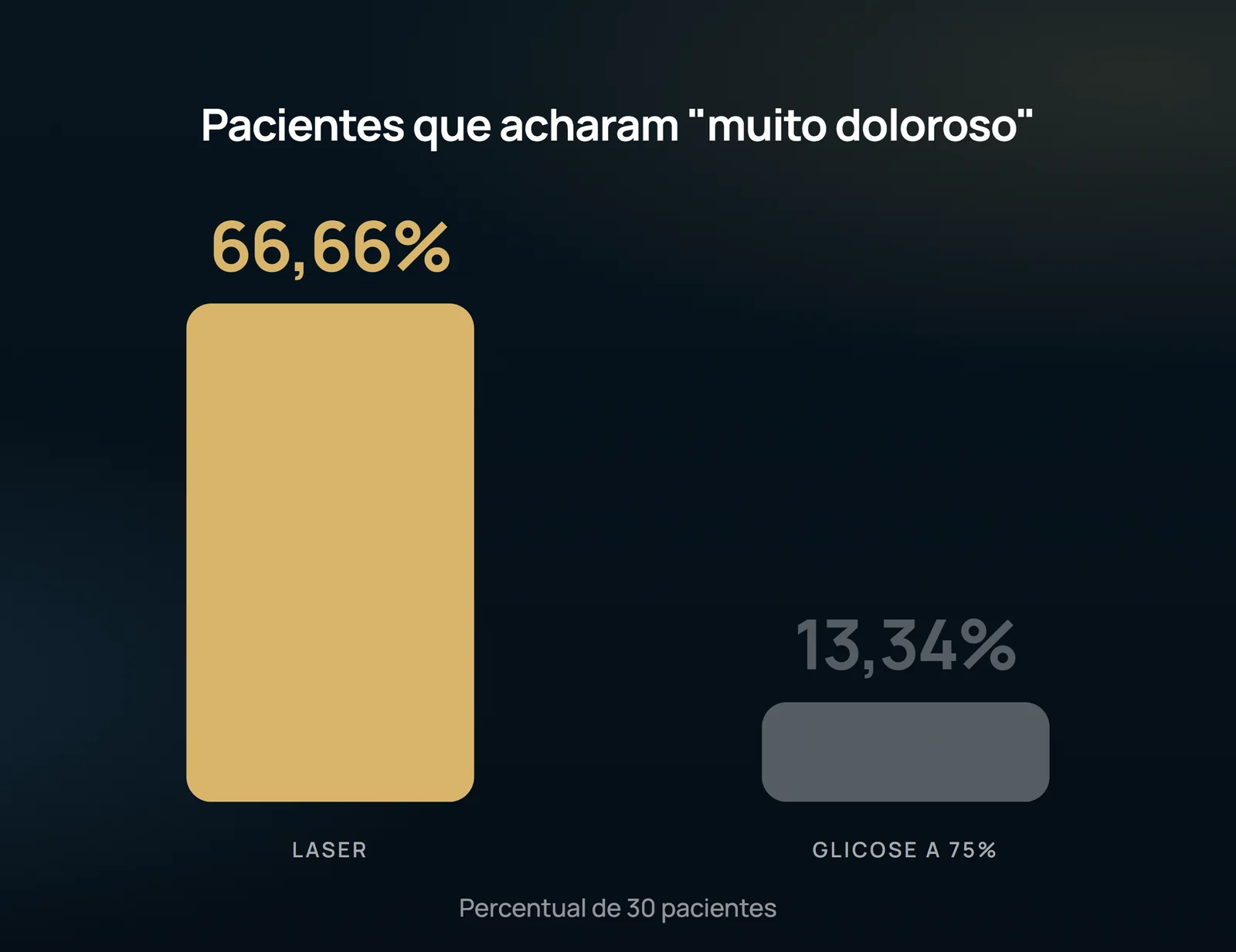
In the evaluation of pain, there was a significant difference between the modalities. They considered the treatment "very painful" 20 patients (66.66%) in laser versus 4 (13.34%) in sclerotherapy; "extremely painful", 3 (10%) in laser and none in sclerotherapy. At the other end, 26 patients (86.66%) classified sclerotherapy as only "lightly painful" versus 7 (22.33%) in laser. The authors attribute this to the fact that they did not use any cooling devices other than the system of the device itself, and remember that smaller spots and longer pulse durations reduce pain.
Regarding clearance in patients' perception, 12 considered laser results good to excellent and 16 said the same of sclerotherapy, with no statistical difference; four classified laser results as poor, and none thus classified sclerotherapy. In satisfaction, 18 were completely satisfied with sclerotherapy against 12 with laser, and four were not satisfied with the laser, a difference that reached significance.
The patients tolerated both modalities well and no serious complications were observed. There was one case of transient post-inflammatory hyperpigmentation related to each modality, and one patient developed a scar in the laser treated area, in a single target site, without compromising the final aesthetic appearance — probably due to previous sun exposure and increased energy absorption by melanin.
What the study does not authorize to state
There are 30 patients in a single center, and photographic control was done 1 week after treatment: the article does not provide any data on recurrence, late matting or results in months. Only CEAP C1 patients were included, so nothing here is transported to the saphenous trunk or varicose veins. The comparison is between a specific device and a specific protocol, and the authors themselves claim that more intense cooling and smaller spots substantially change the reported pain — the measured difference is not a fixed property of Nd:YAG laser. And the text recalls that hyperpigmentation of larger vessels could have been prevented by treating the feeding collateral varices: it is not telangiectasis without treating the reflux that feeds it. The published conclusion is that the two modalities treat equally well, that sclerotherapy costs less and hurts less, and that laser is recommended for patients with telangiectatic matting, needle phobia or allergic reaction to sclerosing — which is an indication, not a promise of outcome.
Content addressed exclusively to health professionals. No individual result is guaranteed: the indication depends on clinical evaluation and imaging examination of each patient (CFM 2,336/2023).