The seated test that reduced indications for varicose vein surgery
A British service changed the order of exams: it placed photoplethysmography as the first objective test of the outpatient clinic before the duplex. With the same surgeon and clinic, the surgery offered dropped from 39% to 24% — and from 51% to 28% in CEAP patients C2.
 View the video lesson
The decision to offer surgery for varicose veins should rest on objective evidence of venous dysfunction, not on the appearance of the limb or the symptom reported. The problem is that objective evidence resides in an exam — the duplex — that no outpatient clinic can offer to every patient who arrives.
Why the symptom is not enough
varicose veins are the most common manifestation of venous disease and affect about 20% of the UK population. In 1999, Bradbury and colleagues at the Edinburgh Vein Study (BMJ) showed a weak correlation between reported symptoms and severity derived from clinical signs, and that surgery did not improve symptoms in most patients. Studies that showed quality of life gain did not have a control group treated conservatively.

The surgical target, therefore, is a subgroup: functional superficial venous reflux. The ideal initial test to separate it needs to be objective, simple, noninvasive, reproducible, sensitive and cheap. The final test needs to be anatomically specific. They are different functions, and the error of using only one of the two has cost in both directions: the isolated manual Doppler is at high risk of losing significant reflux; the universal duplex is not feasible.

What PPG Measures
The transducer emits infrared light in the dermis; the retrospread light depends on the number of red blood cells in the capillary bed and is read by a photodetector as a trace. After rest, the patient performs ten foot dorsiflexions, triggering the calf muscle pump. Venous filling time is the time that the tracing takes to return to 90% of the baseline after the end of the exercise, and is shorter in the limb with reflux. Below 25 s the exam is abnormal, and the exercise is repeated with a venous clamp in the calf or thigh at 60 mmHg to isolate the superficial component.
The design of the study
A prospective observational study, a single vascular consultant, 68 weeks. In the first 22 weeks, the standard protocol (history, clinical examination, manual Doppler or duplex). In the following 46 weeks, the patient was first evaluated by a clinical scientist with a questionnaire and PPG. There were 347 consultations, 239 (69%) of them new referrals: 42 (18%) in CEAP C0/C1, 141 (59%) in C2/C3 and 56 (23%) in C4 to C6.

What Changed in Conduct
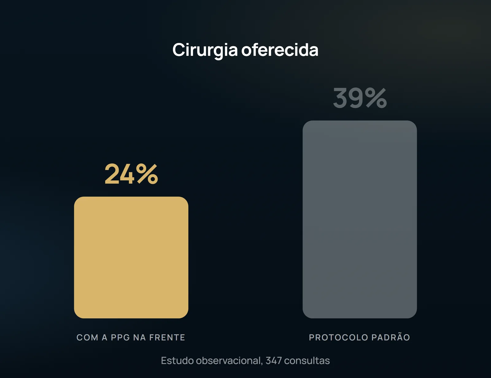
The discharge back to the general practitioner rose from 19% to 29%, and referral to duplex dropped slightly, from 26% to 22%. The change in practice between the two phases was significant (χ2 = 13.3; df = 3; P = 0.004).
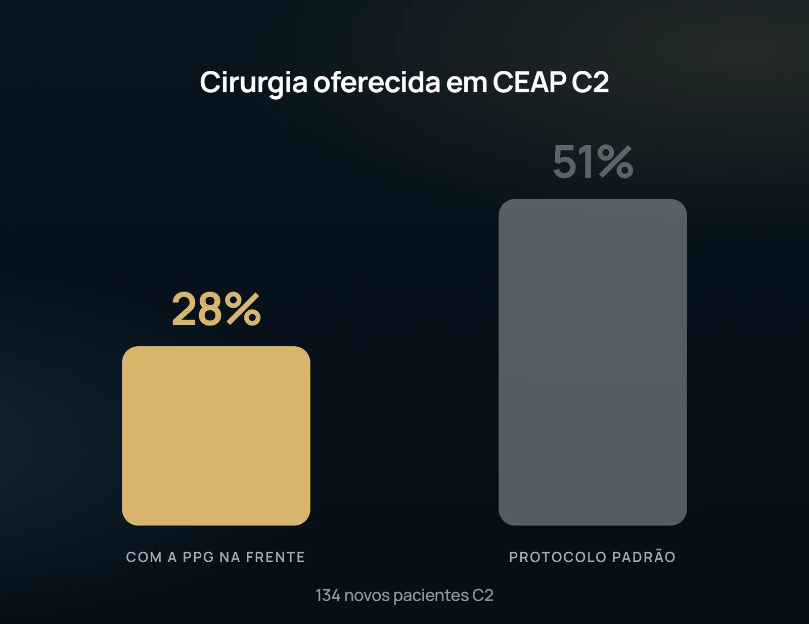
The authors estimate that if surgery was offered only to those with objective evidence of hemodynamic impairment, the surgical load for mild venous disease would fall by about 20%. The economic argument is explicit: In the United Kingdom, community treatment of venous ulcer consumes about ten times the resources of varicose veins surgery — £500 million against £50 million a year.
What the study does not authorize to state
This is an observational study with two consecutive phases, in a single service and with a single consultant surgeon. It is not randomized and does not compare outcomes: it shows that the management has changed, not that patients who no longer underwent surgery have evolved better. The main limitation is recognized by the authors themselves — symptoms were not objectively evaluated in patients who needed surgery, and an additional study would be necessary comparing symptoms and changes in PPG to confirm the hypothesis. There is also a technical limit that the reader needs to carry to the office: PPG measures local venous hemodynamics near the ankle, not the function of the entire limb. Patients with isolated reflux of the anterior vein of the thigh and varicose veins in the thigh may have normal PPG on the ankle.
Content addressed exclusively to health professionals. No individual result is guaranteed: the indication depends on clinical evaluation and imaging examination of each patient (CFM 2,336/2023).