Treating a tortuous tributary with the same fiber used for the saphenous vein
The saphenous inside the sheath is straight and easy to cross. The tributaries outside of it is tortuous — and it is she who decides whether the laser serves. In this series of 173 venous systems, 30% of the largest tributaries systems were excluded due to tortuosity, and in the cases attempted the guide wire J alone was only 43%.
 View the video lesson
Endovenous laser ablation is an established treatment for saphenous reflux. What follows by varying in practice is the fate of the major varicose tributaries associated: ultrasound-guided sclerotherapy or phlebectomy, synchronous or in a second period. This series tested a third pathway — treating the tributaries with the same laser system used in the trunk — and the authors record that they did not find previous reference to this in the literature.
Who came in and who stayed out
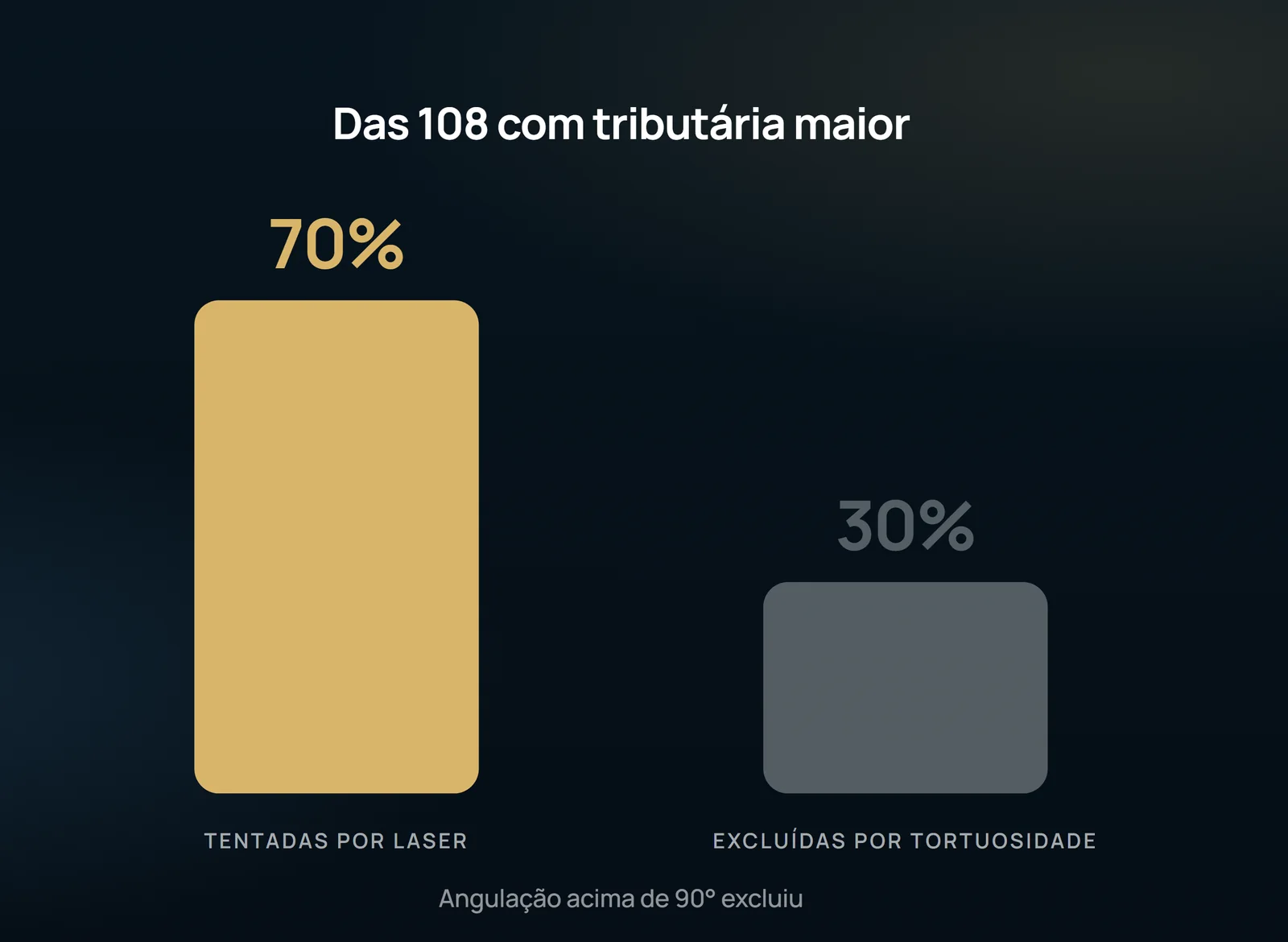
For 12 months, 173 larger venous systems in 95 patients were treated with EVLA by a single professional, in a prospective observational study. In 65 systems (38%) there was no higher tributaries that required treatment. Of the remaining 108, 32 (30%) were considered too tortuous — in general, with segments of more than 90° of angulation — and EVLA was attempted in the remaining 76 (70%), corresponding to 56 patients (36 women, 20 men; 18 to 77 years, median 50).
tributaries were in the anteromedial axis in 60 cases, anterolateral in 13 and posterior in five; above the knee in 25, below in 29 and both in 24. The treated veins were great saphenous vein and tributaries in 51 cases, anterior accessory saphenous and taxal saphenous in 11, great saphenous vein and taxal in five cases, and only tributaries in nine.
Access is the procedure
All were evaluated by duplex with the patient at 45° Trendelenburg reverse on an inclined table. Before the procedure, the veins were marked under ultrasound; 0.2% glyceryl trinitrate ointment at the point of entry and 10% lidocaine gel along the route, under plastic cover, for 30 minutes. The most distal section relatively straight from the tributaries, with diameter ≥4 mm, was punctured with 18G angiography needle under local anesthesia, seeking a "support point" with J guide wire of 0.035" to insert sheath and 5F dilator.
A single puncture was sufficient in 44 systems (58%), two were necessary in 27 (36%) and three in five (7%). Safena and tributaries were treated together as a segment in 49 cases, separately as two segments in 18, and the tributaries alone in nine.
Tumescence and parameters
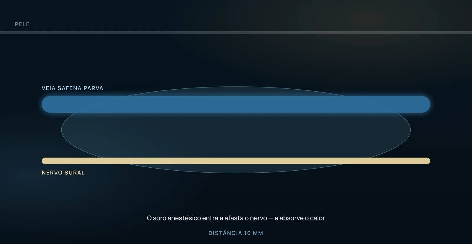
Tumescence was performed with 0.1% xylocaine containing adrenaline at 1/2,000,000, buffered with sodium bicarbonate, by a 9 cm 22G spinal needle and peristaltic pump. Around the saphenous, as usual, with ultrasound control to guide the needle to the saphenous sheath. Around the tributaries, partially under ultrasound and partially by tact, injecting superficially enough to make it impalpable. The volume in the group treated by EVLA of saphenous and tributaries was 55 to 420 mL (median 175 mL).
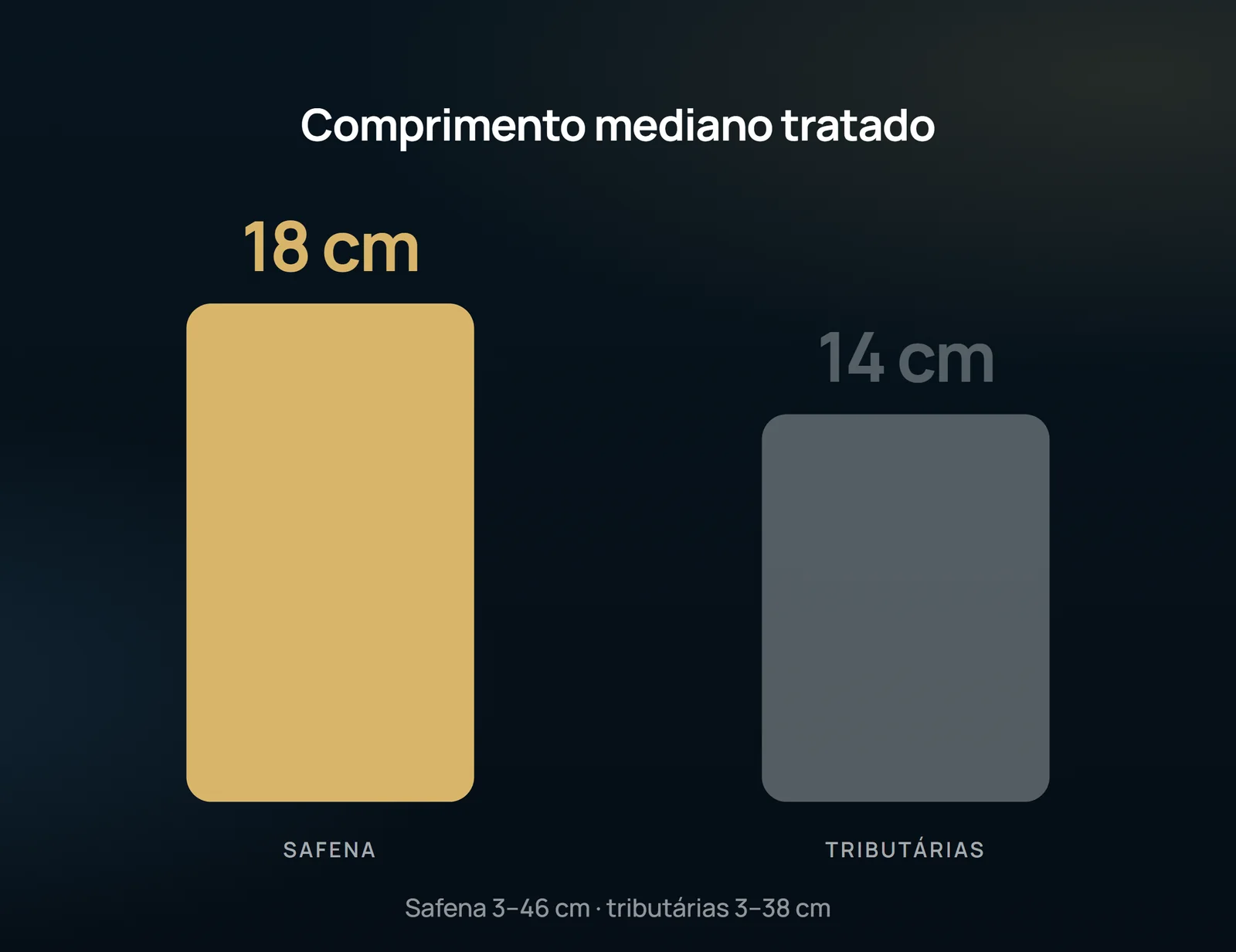
The lengths treated were 3–46 cm in saphenous (median 18 cm) and 3–38 cm in tributaries (median 14 cm). Median diameters were 6 mm in the saphenous and 5 mm in the tributaries. Distal or taxal saphenous veins with diameter less than 3.5 mm were treated by ultrasound-guided sclerotherapy about two weeks later.
Result and safety
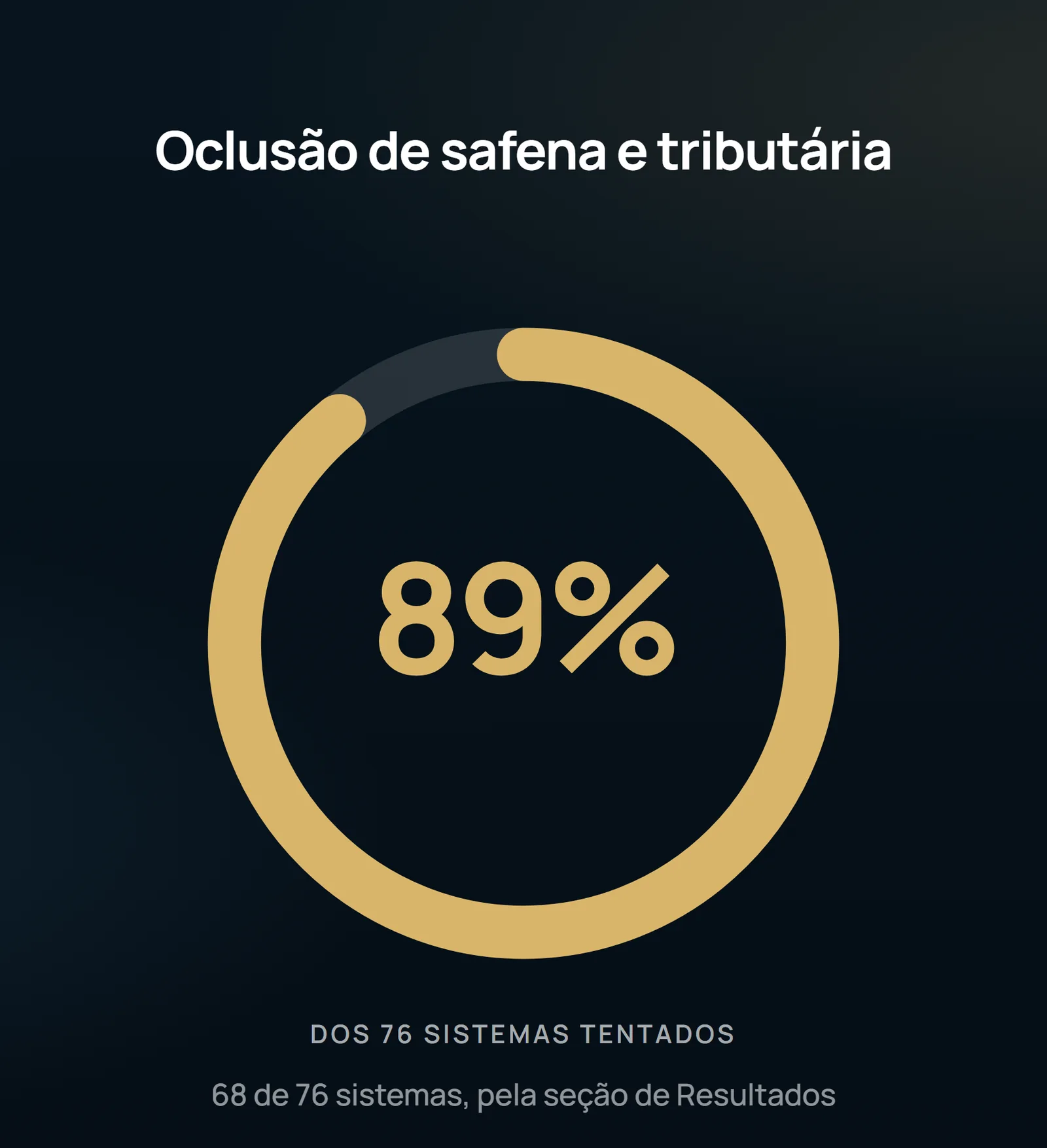
Through the Results section, the technique was successful in the occlusion of both the saphenous and the major tributaries systems in 68 systems (89%); there was failure to pass a wire through the tributaries in seven patients and failure to control the saphenous in one. In the discussion, the authors record that the wire could be manipulated by considerable tributaries lengths in more than 80% of the selected venous segments. Ultrasonographic surveillance in the following days confirmed saphenous and tributaries-related occlusion after successful treatment.
There was hematoma along the tributaries in all patients, resolved in about four weeks without residual pigmentation. There was no phlebitis in any patient, and the tributaries dollars were barely palpable. Two patients felt pain at the passage of the wire. There was no skin damage, saphenous or sural nerve injury, or other early complications, and there were no late sequelae such as persistent hardening or fat atrophy.
What the study does not authorize to state
This is a preliminary, observational communication conducted by a single professional, and the authors explicitly state that no attempt was made to compare the results with the treatment of tributaries by guided sclerotherapy or phlebectomy. There is therefore no basis for asserting superiority over either of the two. The follow-up is fragmented and short: eight systems reviewed in the first week, 15 in six weeks, 35 in six months and only 18 in 12 months — which does not allow conclusion on late recanalization or recurrence. The criterion for selection by tortuosity is clinical and not measured judgment: the authors themselves claim that it was impossible to quantify the degrees of tortuosity. The equipment is specific (the 1500 nm system), and the parameters described are valid for it. Finally, the summary and section of Results of the article differ in some numbers (78 versus 76 systems treated; 83% versus 89% success): the values used here are those of the Results section.
Content addressed exclusively to health professionals. No individual result is guaranteed: the indication depends on clinical evaluation and imaging examination of each patient (CFM 2,336/2023).