Endovenous laser in the interventional suite: access, contrast and 12 watts
The 2007 Korean series is remembered for the failure of direct laser in tributaries. Before that, however, it describes a route of access to the saphenous that today is hardly used: fluoroscopy, contrast venography, advanced 0.035 wire beyond the junction and power of 10 to 12 W in the magna. The route works — her price has never been measured.
 View the video lesson
Everyone cites this article by the negative conclusion: direct ablation of branch varicosities does not replace phlebectomy or sclerotherapy. But the Materials and Methods section describes something else — a saphenous endolaser performed by interventional radiologist on the fluoroscopy table with contrast venography. For those who have already lost distal track on a magna, this route is worth reading.
Who entered and how it was diagnosed
From July to August 2005, 22 patients with CEAP C2-C4 varicose veins were referred to the interventional radiologist and submitted to duplex. Seventeen entered the prospective study — 21 limbs, 18 saphenous magnas and 3 parva, male proportion: female 7:10, mean age 50.1 years (range 24 to 62). The reflux was defined as a reversal of saphenous flow for more than 0.5 seconds after the release of compression in standing and during the supine Valsalva. Deep venous thrombosis, non-palpable pedious pulse, inability to walk and allergy to sclerosing were among the exclusion criteria.
The access route, step by step
The dumb saphenous was accessed in the lower calf with a Jelco 22 gauge needle. The magna had three addresses: ankle or knee, also with Jelco 22, or groin, with a 21 gauge micropuncture needle, when the distal access could not be reached. The ankle magna was punctured directly; dumb, knee and groin were punctured under sonographic guidance. Then a 0.018 inch guide wire and a 4 or 5 Fr microsound were entered under fluoroscopy, and a contrast-enhanced venogram verified the course of saphenous and its tributaries.
The next step is what is most common in current practice: a 0.035-inch guide wire was advanced anterogradely beyond the saphenofemoral or saphenopopliteal junction, to the iliac or popliteal vein, under ultrasound guidance — and, in cases accessed by the groin, advanced retrogradely beyond the knee saphenous. Over it went a 5 Fr guide catheter, and inside the catheter the 600 μm naked-tip fiber and 980 nm, advanced to the second mark, projecting 3 cm of fiber out of the tip. The final position, 1 to 2 cm from the junction, was confirmed by ultrasound.

Tumescence, power and postoperative
The tumescent solution — from 100 to 250 mL of 0.05% lidocaine — was delivered by percutaneous needle, under ultrasound, along the saphenous course and inside the fascial envelope. The energy was applied in continuous mode: 8 to 10 W in the small saphenous vein and 10 to 12 W in the great saphenous, with an average removal of approximately 5 mm per second, catheter and fiber pulled together. In the postoperative period, immediate discharge, graded thigh compression compression stocking class II (30 to 40 mmHg) for at least 1 month, immediate ambulation and prescription of analgesic and antibiotic for 3 days.
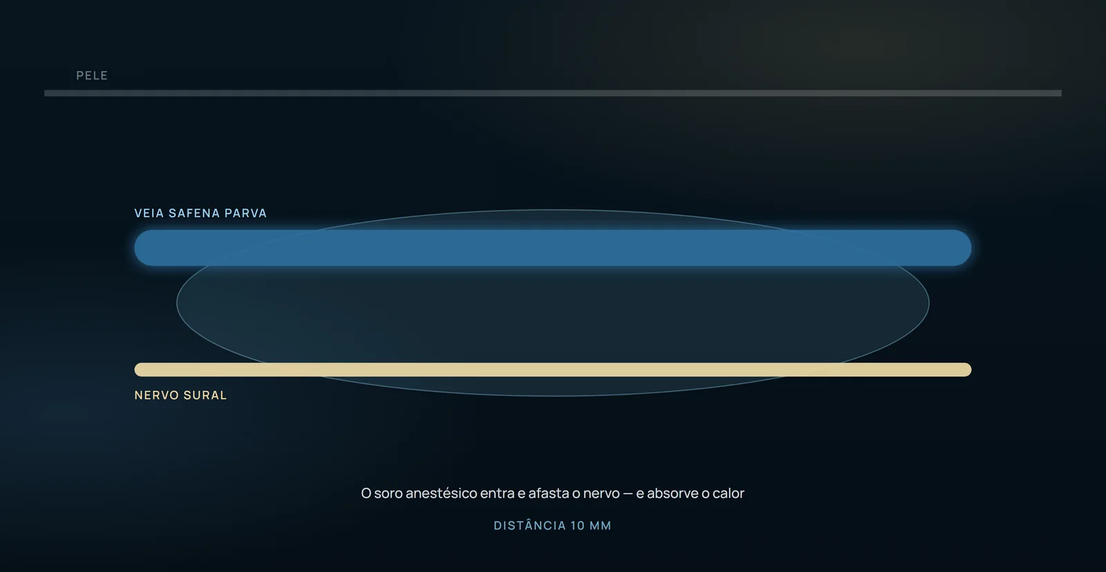
What the log delivered
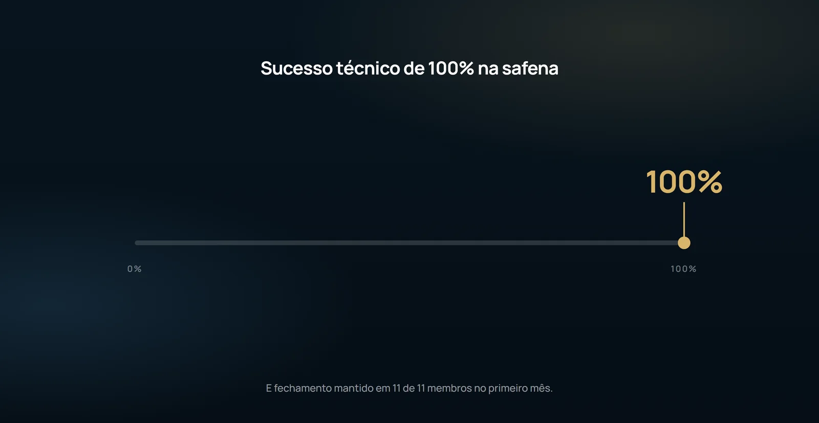
That number needs a scale. The article's own discussion records that the saphenous occlusion rates reported in the literature, with wavelengths of 810, 940 and 980 nm, range from 87.9% to 100% at the end of follow-up. Closing the trunk is not the route differential. And closing the trunk also does not close the case: in the Disselhoff series cited by the authors, 20 limbs (21.5%) maintained persistent varicosities of the superficial accessory vein of the magna and needed additional treatment — compressive sclerotherapy in 16.7% and phlebectomy in 5.6%.
What the study does not authorize to state
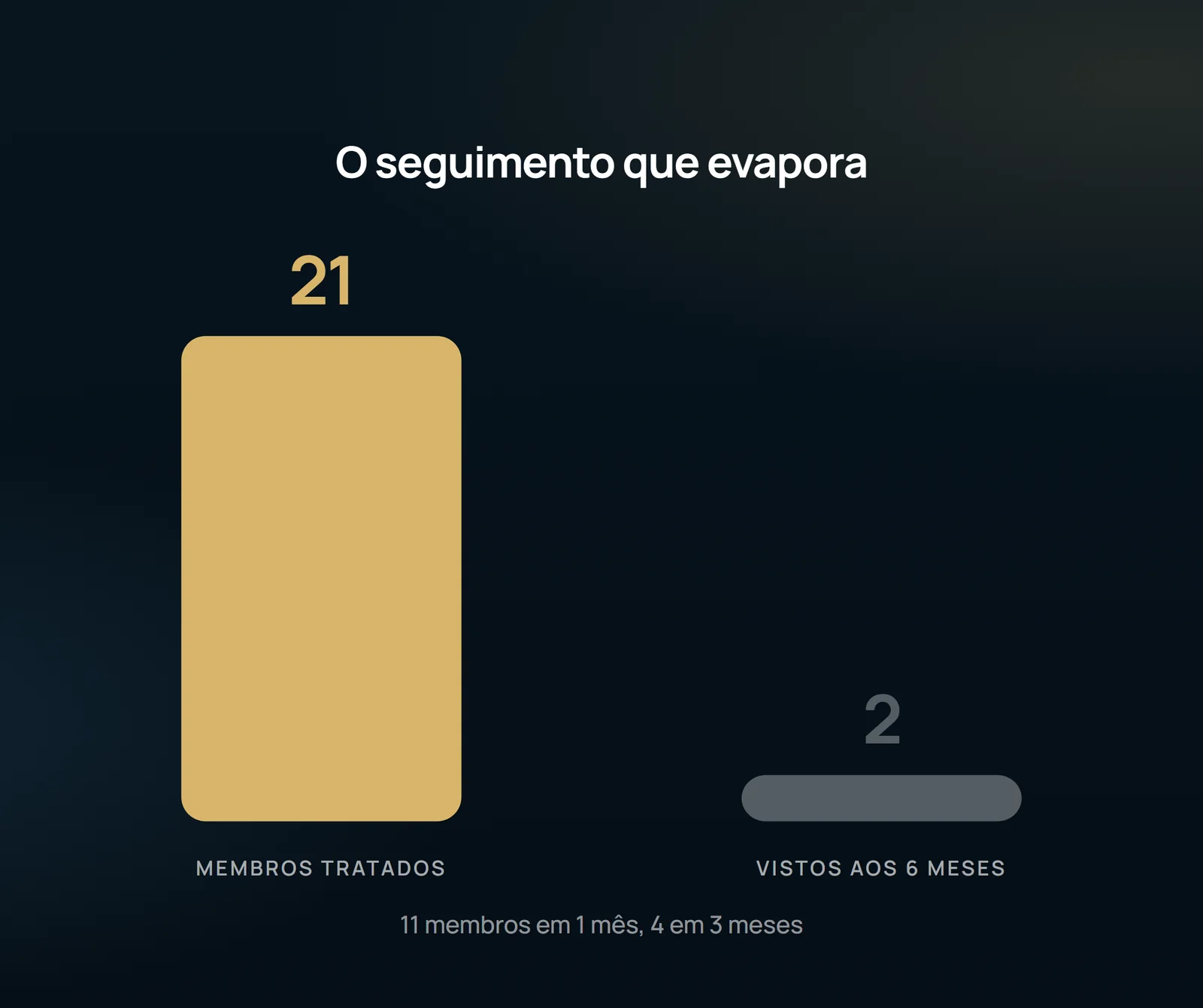
The series cannot establish durability: 21 limbs were treated, follow-up was available for 11, with 4 limbs assessed at 3 months and 2 at 6 months. It also cannot establish that fluoroscopic access is better than ultrasound-guided access: there was no comparator group or randomization, recruitment lasted 2 months at a single center with 17 patients, and the treating surgeon and radiologist assessed outcomes without blinding. Important information is missing for a procedure using fluoroscopy and iodinated contrast: no radiation dose, contrast volume or renal-function data were reported. The access method is described, but its associated burden is not. A vascular surgeon's ultrasound assessment determines the appropriate access for each leg.
Content addressed exclusively to health professionals. No individual result is guaranteed: the indication depends on clinical evaluation and imaging examination of each patient (CFM 2,336/2023).