Foam first, laser second — with a window of 1 to 3 minutes between them
A randomized trial with blind evaluation followed 517 legs treated with polydocanol microfoam followed by Nd:YAG laser, against 79 treated only with foam, for three years. The sequence — and waiting between the two steps — is what defines the method. And she has a cost in pain.
 View the video lesson
Sclerotherapy remains the gold standard for telangiectasis and reticular veins with less than 4 mm. The bottleneck is not the efficacy per vessel, it is the area: when varicosity covers most of the two legs, the dose limit per session imposes a high number of sessions and maintenance treatments. That's the problem the combined method tries to solve.
The design of the test
Randomized clinical trial, controlled by polydocanol, with blind evaluation. We recruited 320 patients, all women, 19 to 72 years old (mean 46.3), Fitzpatrick II to IV phototypes, with more than 20% of the surface of the affected leg and without previous treatment. Deep system reflux, confirmed by Doppler, was an exclusion criterion. Randomization by sealed envelope allocated 550 legs to combined treatment and 90 to polydocanol alone; with losses, the analysis was in 517 and 79 legs. Three blind doctors classified the photographs (kappa 0.86 and 0.81 against the first evaluator).
The technique, step by step
The microfoam was prepared using the Tessari technique: 2 mL of 0.3% polydocanol to 8 mL of air, alternated between two syringes 15 times. The injection used a 2 mL syringe and 30G needle, with a maximum of 20 mL in both legs added — usually less than 10 mL per leg. The injection bleaches the vessels; it follows a wait of 1 to 3 minutes until it recovers a soft pink color, and only then is the laser applied throughout the route.
A long-pulsed 1064 nm Nd:YAG laser was used, with a 2 to 5 mm spot selected for vessel diameter and simultaneous skin cooling through a glass chamber with airflow at 4 °C. Pulse energy was 9.42 J with the 2 mm spot and 11.77 J with the 5 mm spot; pulse duration was 30 ms for classes I and II and 50 ms for class III, with repetition rates of 5 Hz and 2 Hz, respectively. Each leg took 20 to 25 minutes, and treating both whole legs took less than 1 hour. Two sessions were performed 3 weeks apart, followed by compression stockings for 2 weeks.
The result at three years
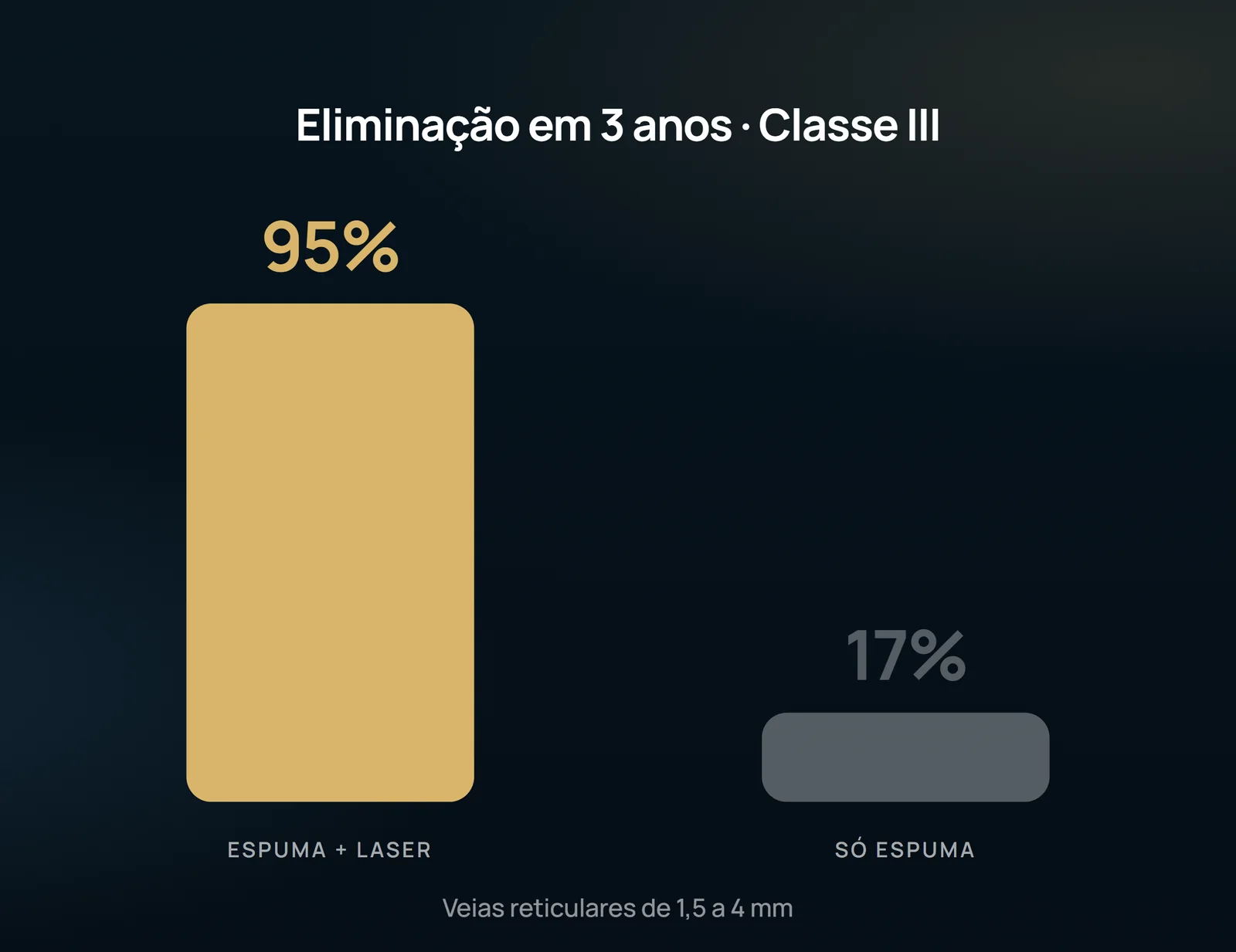
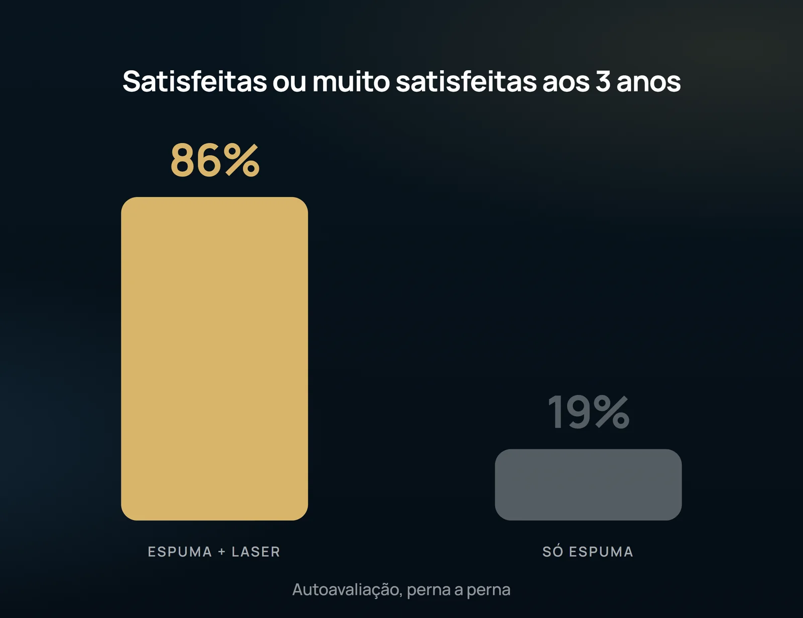
The pattern is repeated in the other classes: 89% against 15% in Class I (red vessels less than 0.5 mm) and 94% against 18% in Class II (vinectasis from 0.5 to 1.5 mm). The difference was significant at all times and all classes (p < 0.001). In the polydocanol alone group, mean elimination rates at three years were below 20% and in some cases varicose veins worsened. In self-assessment, 86% of the legs in the combined group were classified as satisfied or very satisfied at three years, compared to 19% in the polydocanol group alone.
The Cost: Pain

There were no withdrawals due to pain, and the authors attribute tolerance to the dosage applied, cooling systems and a possible anesthetic effect of polydocanol itself. As for adverse effects, 7.73% of the legs in the combined group showed some change at three months — hyperpigmentation (5.02%), hypopigmentation (1.16%), blisters (1.16%) and matting (1.54%). All resolved, except for four cases of hypopigmentation (0.77%) that persisted until the third year.
What the study does not authorize to state
The elimination scale used does not discriminate well between 4 points (80%) and 5 points (100%) — the authors themselves record this limitation, and a note 5 only indicates that the result is closer to 100% than 80%. The outcome is photographic and subjective, classified by blind evaluators, not an objective measure of occlusion. The population consists only of women, from a single center, with reflux of the deep system excluded by Doppler: nothing here applies to insufficient saphenous trunk or vessel above 4 mm. The authors also note that it is not determined whether new venectasis will form in the long term, nor whether the method has preventive value. It is evidence of efficacy and durability in three years for a specific patient profile — no guarantee of outcome, and the indication remains individual.
Content addressed exclusively to health professionals. No individual result is guaranteed: the indication depends on clinical evaluation and imaging examination of each patient (CFM 2,336/2023).