3 ms versus 60 ms: constrict the vessel or simply thrombose it?
Spot and fluence are adjusted in every session; pulse duration almost always stays in the factory pattern. A study with six durations tested in the same patient shows what this parameter decides: whether the vessel contracts or just thrombi.
 View the video lesson
Spot, pulse duration and fluence are the three determinants of laser treatment of leg veins — but until this study, the optimal pulse duration for Nd:YAG had not been established in humans. The design is simple and elegant: in each patient, test sites with 3, 20, 40, 60, 80 and 100 ms, in vessels of approximately equal size, and then up to 20 vessels treated with the duration that had achieved the best occlusion with the lowest adverse effect.
Who entered the study
Twenty-one patients, all women, aged 27 to 82 years (mean 51 years), with Fitzpatrick I to IV phototypes distributed in 9.5%, 57.2%, 23.8% and 9.5%. Eighteen completed: one abandoned due to hyperpigmentation after the test sites and two were lost in the follow-up. The treated vessels had diameters from 0.1 to 1.6 mm, with a mean of 0.8 mm, and the thigh was the most frequent site, followed by the popliteal fossa. No adjuvant treatment — or compression stockings were used.
What the short pulse does differently
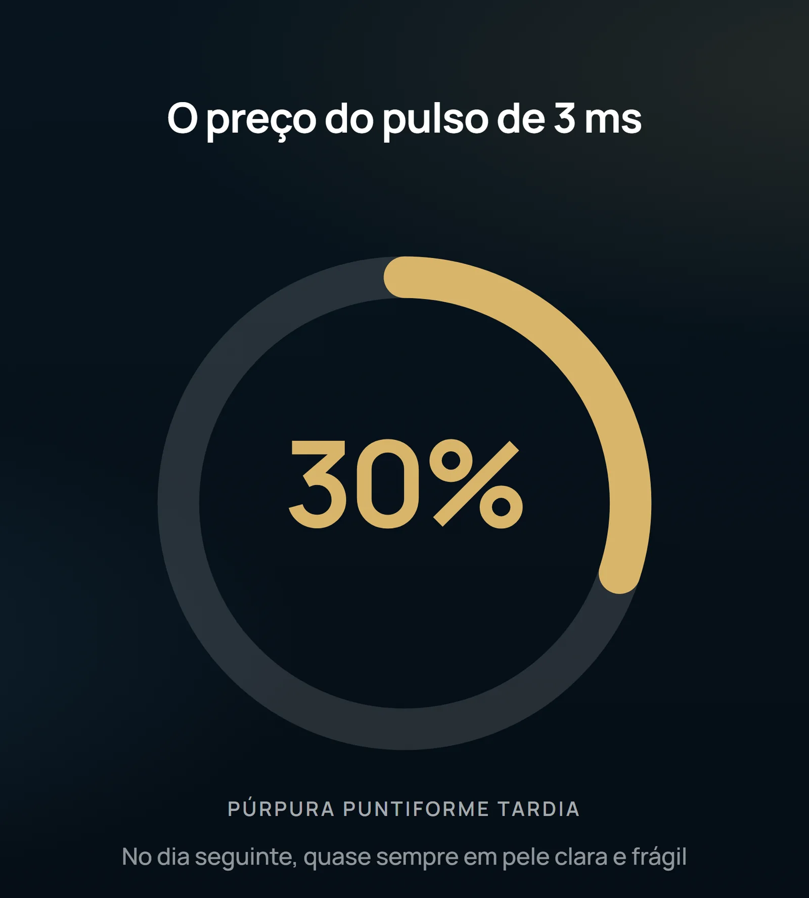
At 3 ms, in 80% of cases the vessel bluished immediately after irradiation — coagulation and thrombus formation — but the contraction was modest or nonexistent. It was the only duration whose immediate reaction differed significantly from all the others. The same pulse produced late punctiform purpura one day after treatment, in 30% of patients, almost always those with clear and fragile skin. In post-hoc comparisons, the difference in purpura and edema was only significant between 3 and 40 ms and between 3 and 60 ms.
Stenosis is preferable to thrombosis
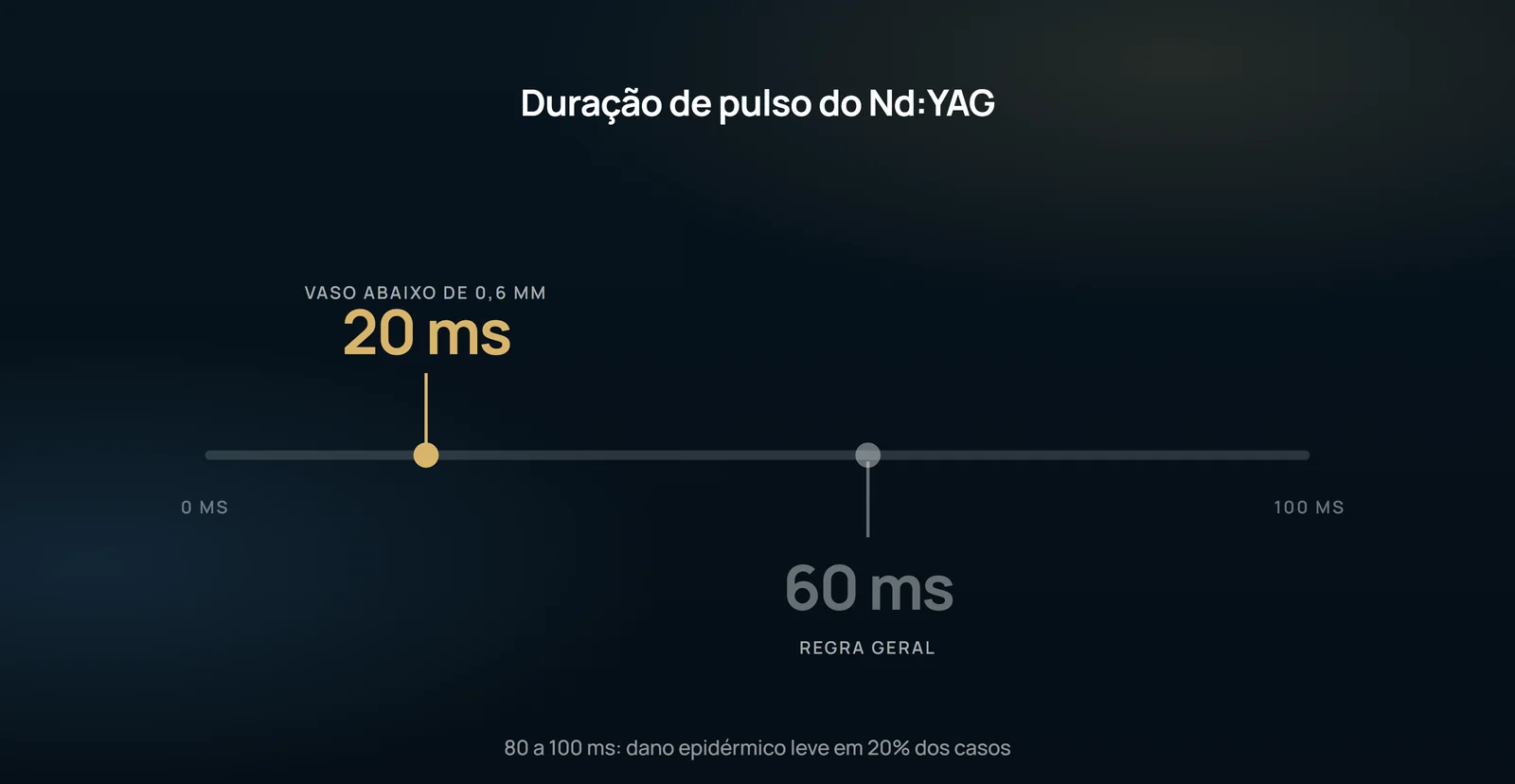
Most vessels showed more complete stenosis, with less purpura and less edema, in the durations of 40 to 60 ms; 60 ms was the optimal pulse in most patients and 40 ms was comparable in most of them. Pulses of 80 to 100 ms had similar performance, but required slightly higher fluence and, in larger veins, produced mild epidermal damage in 20% of cases, presumably by retrograde heating of the epidermis from the vessel. The mechanical explanation is in the histology: with the same parameters (1.5 mm, 0.8 mm vessel, 500 J/cm2), the pulse of 3 ms showed denatured blood clot filling the lumen, while 60 ms showed contraction of the lumen with endothelium hyalinization and perivascular collagen. The ratio between contraction and thrombosis increases with pulse duration — and stenosis produces fewer side effects.
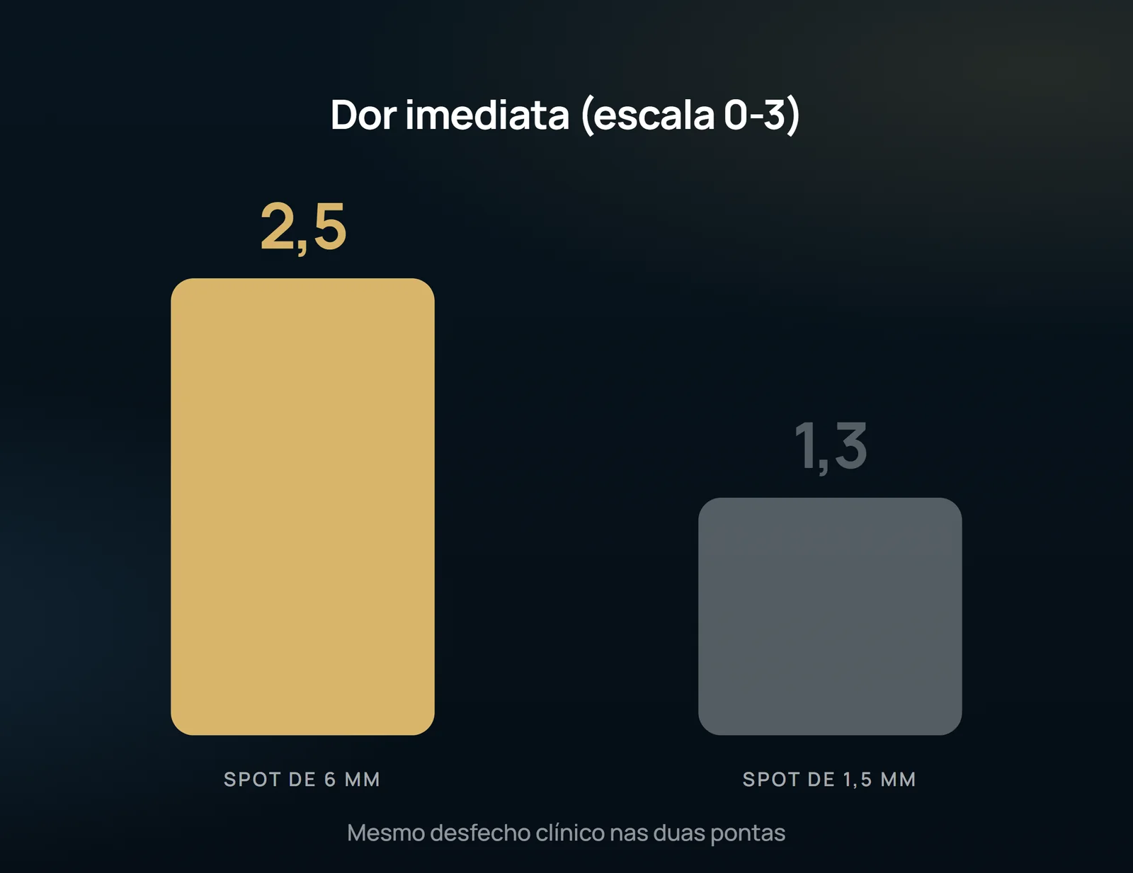
There is a relevant exception to the protocol: in very small vessels (< 0.6 mm), the pulse of 20 ms resulted in better closure than that of 60 ms, contrary to the general finding. And the required fluence depends on the spot: 80 to 200 J/cm2 for 6 mm, 200 to 300 J/cm2 for 3 mm and 280 to 580 J/cm2 for 1.5 mm — roughly, about 1.7 times the fluence at each spot reduction for the same immediate reaction. The spot, in turn, is the main predictor of pain: with equivalent clinical outcomes, the mean scores were 2.5 with 6 mm, 2.1 with 3 mm and 1.3 with 1.5 mm. Cooled air cooling relieved pain more than cryogen spray, with no difference in clinical outcome.
What the study does not authorize to state
There are 18 patients who completed the protocol, all women of phototypes I to IV — phototypes V and VI were exclusion criteria, as well as concomitant venous stasis, anticoagulation, thromboembolic condition and pregnancy. There is no control group or comparison with sclerotherapy, which the article itself recognizes as the gold standard. The follow-up was 16 weeks, with the clearance outcome judged in photographs, and the kappa of 0.445 indicates only moderate agreement among the evaluators. None of this allows you to design long-term durability or relapse rate.
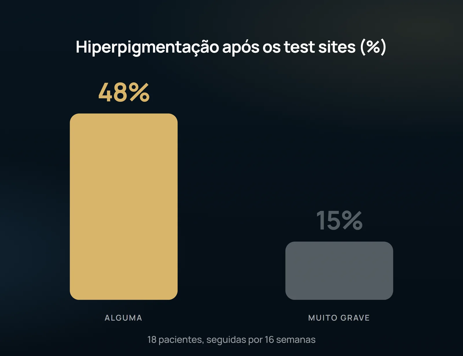
Adverse effects should also not be minimized: hyperpigmentation occurred in 48% of patients after the test sites and was classified as very severe in 15%; in severe cases, resolution was only about 50% in 16 weeks, and very mild hyperpigmentation persisted even after four additional months of follow-up outside the protocol. One patient developed a pinpoint blister by accidental pulse stacking, resulting in a slightly perceptible hypopigmented scar. The final recommendation of the authors is technical and limited to the studied range: for vessels from 0.1 to 1.6 mm, optimal occlusion is obtained with pulse durations of 20 to 60 ms, using the lowest fluence and the smallest spot that achieve complete immediate stenosis. This is a reference parameter, not a promise of results: the indication and protocol depend on the individual evaluation made by vascular surgeon.
Content addressed exclusively to health professionals. No individual result is guaranteed: the indication depends on clinical evaluation and imaging examination of each patient (CFM 2,336/2023).