In the office, anesthesia is part of the technique
Sclerotherapy, outpatient phlebectomy and percutaneous venous ablation fit in the office. What separates a tolerable procedure from a bad procedure is not the device — it is the dilutions, the anesthetic mixture, the order of the needles and the method of attaching the tape.
 View the video lesson
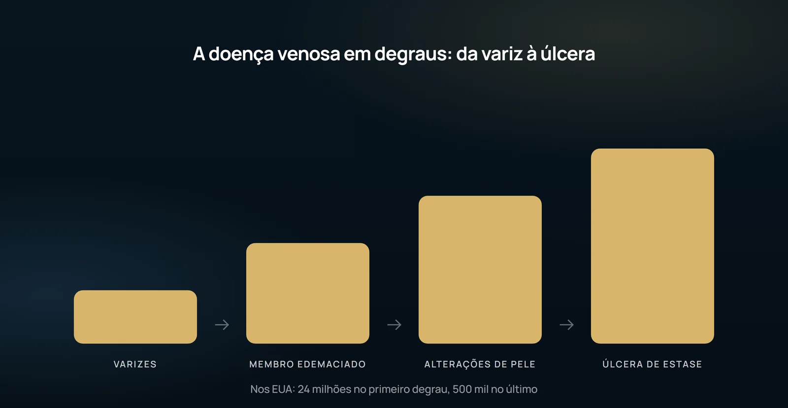
Chronic venous disease is 10 times more prevalent than arterial disease in the United States: approximately 24 million Americans have varicose veins, 6 million have limb edema, 1 million have chronic skin changes and 500,000 have stasis ulcers. This volume supports moving venous procedures from the operating room to the office — making anesthesia a central part of the technique rather than an accessory detail.
Enlightening: three families and dilution
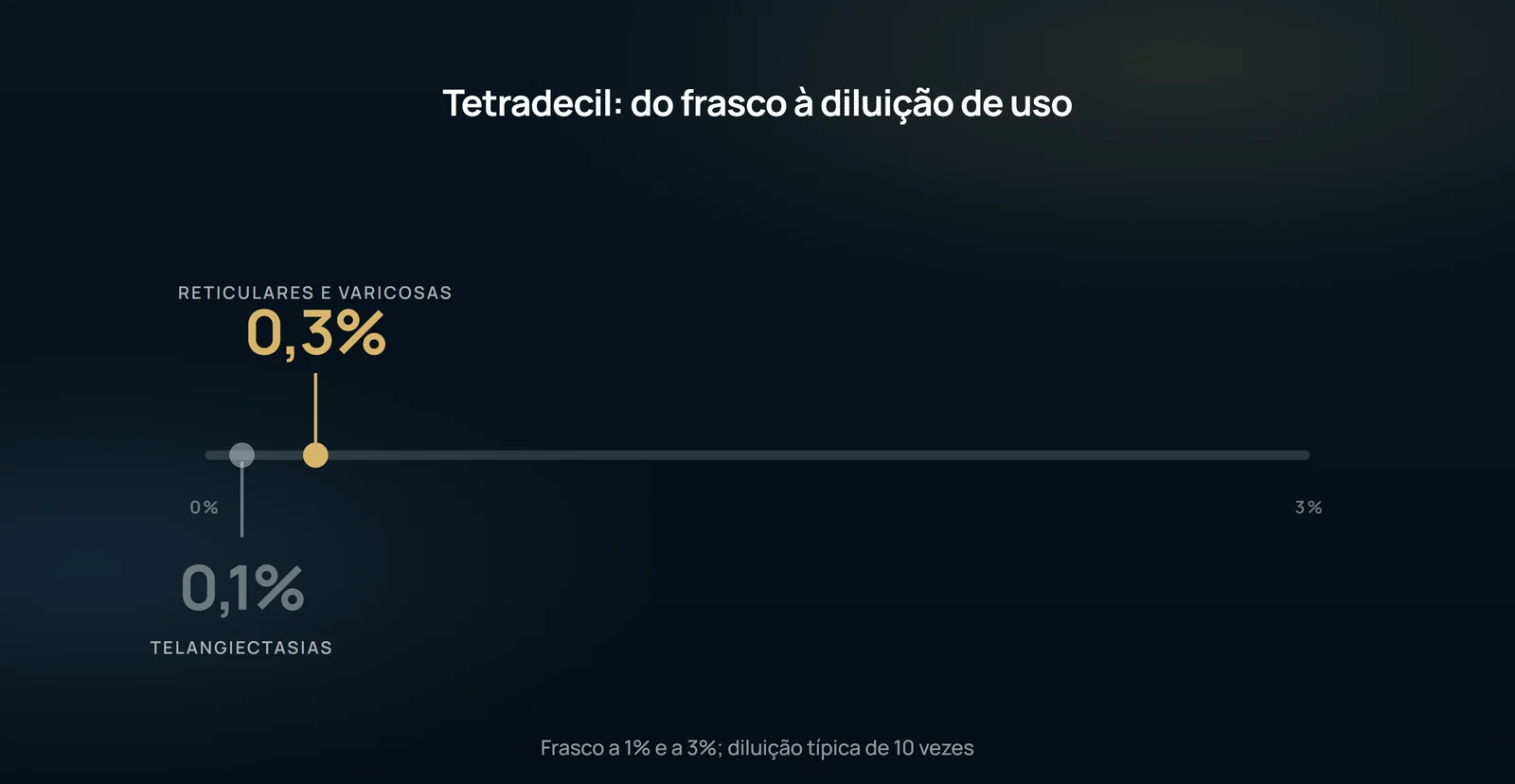
There are three categories of sclerosing material: detergents (sodium morruitate, ethanolamine oleate, polydocanol, sodium tetradecyl sulfate), osmotic agents (hypertonic salina, hypertonic glucose, sodium salicylate) and chemical corrosive agents (sodium and potassium iodide, chromoglycerin, absolute alcohol). Sodium tetradecyl sulfate is the one approved by the FDA for aqueous injection of telangiectasis, reticular veins and varicose veins; it comes in vials of 2 mL, 1% and 3%. The dilutions are typically 10 times: 0.1% for telangiectasis and 0.3% for reticular and varicose veins. Listed contraindications include use of oral contraceptives, pregnancy, a strong history of allergy and morbid obesity — the latter because adequate compression is not achieved.
The foam was reintroduced by Cabrerra in the early 1990s and had its technique optimized by Tessari in 2000. The tap light is closed slowly as the mixture continues to favor the formation of microbubbles. The point of attention is systemic: transient ischemic attack and stroke have been reported after foam sclerotherapy in patients with patent foramen ovale. The described approach is to position the patient in Trendelenburg to prevent air from travelling centrally; ultrasound guidance helps, because it allows you to see the microbubbles and manually contain the passage into deep circulation.
Ambulatory phlebectomy: mixing and the millimeter
The patient should remain standing for approximately 5 minutes to ingorge as much as possible the varicosity before marking with a permanent pen; only then will he go to Trendelenburg from 20° to 30°. The anesthetic mixture is explicit: 1% lidocaine in equal parts with 0.5% bupivacaine for immediate onset and long duration, with 8.4% sodium bicarbonate added as 10% of the total to reduce burning. If more than 50 mL are required, tumescence is used: 50 mL of the lidocaine/bupivacaine mixture in 450 mL of 0.9% saline. For phlebectomy, the local anesthetic goes without epinephrine.
Trapped and brought by the incision, the vein is released from the surrounding tissue without the incision being stretched; taking varicosity at the base and applying a firm swing movement brings more varix closer. The avulsion occurs when nothing else can be released without increasing the incision. The closure is with benzoim and sterile compression stocking-inch paper ribbons, applied transversely and without tension — if there is tension, bubble form, which can lead to long-term skin discolouration. The following is a multilayer elastic bandage, and the patient walks after the procedure.
Ablation: the eye sign and order of needles
The great saphenous vein is the most ablated vein, but not the only one: the anterolateral branch of the thigh, the small saphenous vein and the Giacomini vein are also treated safely. Ultrasonographic orientation should be used at all times when tumescence is applied. In the transverse image, the saphenous fascia around the vein often mimics the appearance of an "eye": the 18-G needle is guided into this fascia and, when injected, the liquid should completely surround the wall of the vein, pushing the fascia away. Made correctly, the liquid naturally dissects along the saphenous and minimizes the number of injections required.
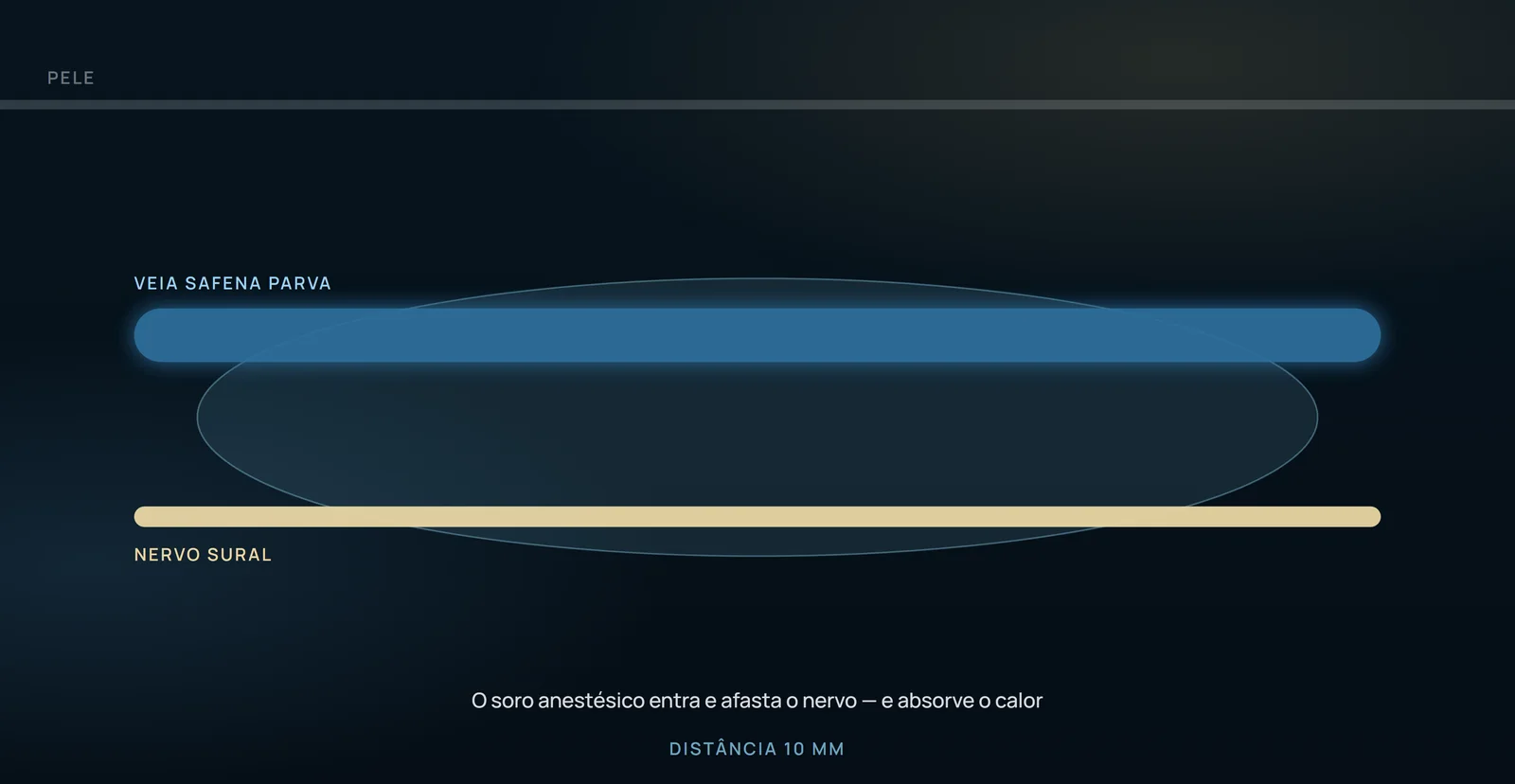
The maneuver that makes this painless is sequence: quickly map the great saphenous at ultrasound, are marked with a permanent pen equidistant points where the tumescent fluid will be injected and, with a 27 G needle, small skin buttons are made with 1% lidocaine at each point. These areas are then punctured with the 18 G larger needle. Automatic injectors with smaller needles are also available and may be useful when several ablations will be made on the same day.
What this article does not allow to say
This is a perspective article presented at a postgraduate course in September 2006. It reports no original series, randomization, comparator group or success, occlusion or recurrence rate. It cannot establish that one technique is superior or promise an outcome to a patient. Its only quantitative outcome data are charts of consultation and procedure volumes before and after establishing vein centers in Nashville (population about 1 million) and Springfield (about 110,000), across two consecutive 3-year periods. These are service-organization data, not clinical outcomes. Sclerotherapy complications are listed without frequencies: vasovagal, allergic or anaphylactic reactions, toxic reactions and hemolysis, skin necrosis and ulceration, venous thrombosis, arterial injection, nerve injury, dyschromia and telangiectatic matting. The article should be read as a practical guide from an experienced service.
Content addressed exclusively to health professionals. No individual result is guaranteed: the indication depends on clinical evaluation and imaging examination of each patient (CFM 2,336/2023).