The forehead vein drains into the cavernous sinus
The dilated frontal vein looks like a surface problem. It is not: part of its drainage follows through the ophthalmic veins to the cavernous sinus. A Brazilian case report describes the anatomy, risk and three maneuvers required by the technique.
 View the video lesson
The veins of the frontal region of the face can exaggerate in crying, smiling, stress, physical exertion and lowering the head — and the thinning of the skin and atrophy of the subcutaneous with age worsen the appearance. What makes this complaint a vascular problem, not an aesthetic one, is the fate of the blood that passes there.
The Anatomy of Drainage
The frontal veins — right supratrochlear (RSV) and left suprachlear (LSV) — originate in the forehead and communicate with the frontal branches of the superficial temporal vein. They converge forming a single trunk that passes through the midline of the forehead and communicate at the root of the nose through a transverse branch, the nasal arch. They separate in the nose and join the supraorbital vein to form the angular vein. Angular drainage is, in part, directed by the ophthalmic veins superior and inferior to the cavernous sinus; the other route is the facial vein, which drains into the internal jugular.
The risk is named bluntly in the article: inadvertent administration of the sclerosing agent can lead to iatrogenic aseptic thrombosis of the cavernous sinus, with a picture that can combine swelling, vascular engorgement, orbital pulsation and exophthalmia. Therefore, before indicating treatment, the authors study anatomy and flow with Doppler ultrasound and increased reality, inducing a change of calibre in the office with the patient in frontal decubitus and Trendelenburg position.
The case: the isolated laser did not resolve
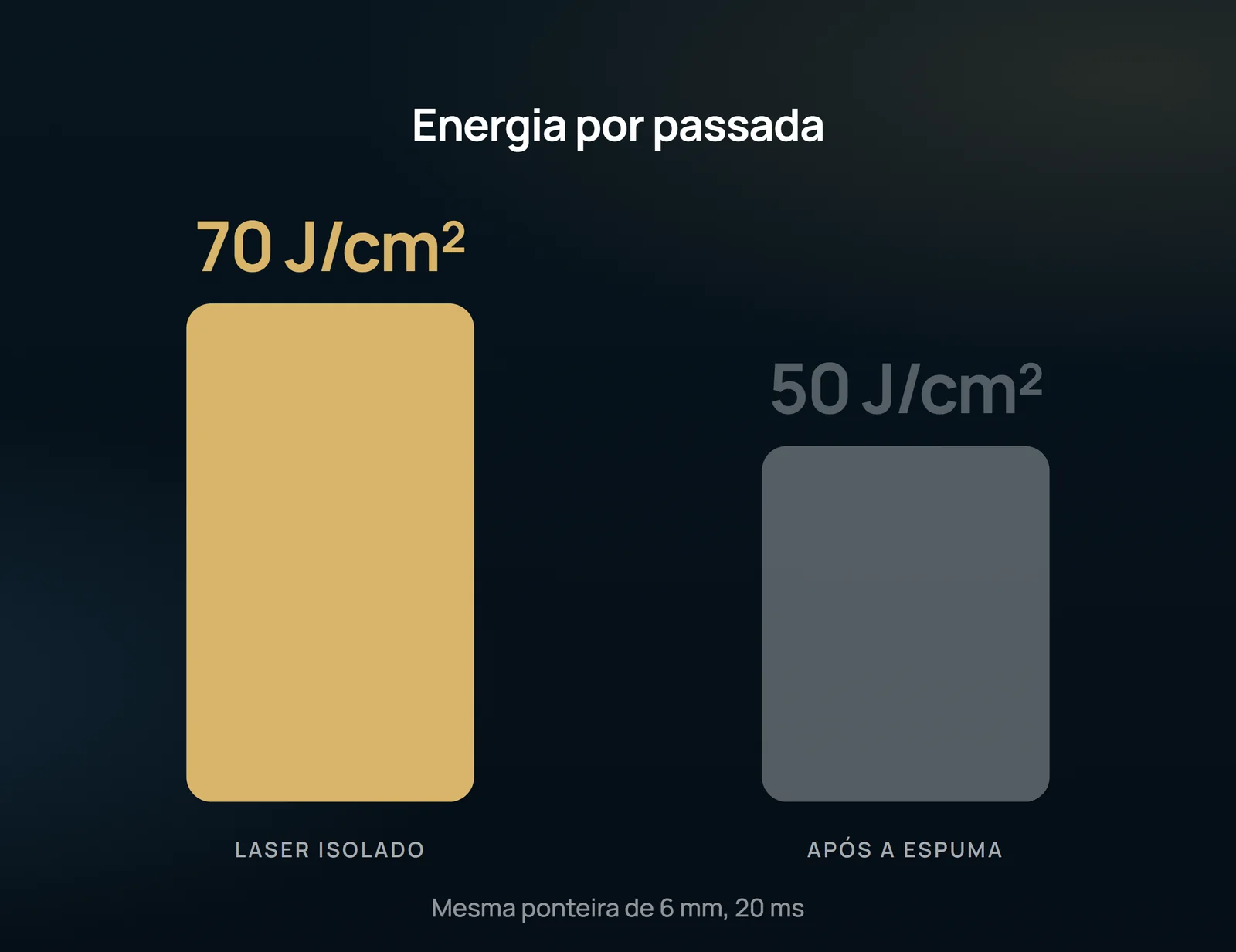
A 43-year-old dancer with no comorbidities was seen in February 2022 due to non-esthetic discomfort with the vessels of the forehead, with significant dilation after physical effort. The initial approach was Nd:YAG 1064 nm of long pulse isolated: 2 sessions with a 15-day interval, 6 mm tip, 20 ms of pulse time and 60 to 70 J/cm2, with 3 passes throughout each session. In the third visit, there was no regression of the frontal vein.

The combined technique followed three steps that the report describes as deliberate. First position: patient in frontal decubitus, Trendelenburg, with puncture of each supratrochlear made as close to the scalp as possible. Then, during the injection, manual compression in the glabella region, with the stated objective of closing the drainage to the angular vein. Only then the laser QS Nd:YAG 1064 nm, with 3 passes throughout the extension, 50 J/cm2, 20 ms and spot of 6 mm — and compressive dressing in the path of veins at the end. Within 15 days, the left frontal vein was no longer visible either during sports practice or after Trendelenburg maneuver; the right required a new session, with the same 0.3 mL and laser from 40 to 50 J/cm2.
Why Nd:YAG has a limit here
The Nd:YAG 1064 nm is described as the most versatile laser for facial vessels, useful in telangiectasias and reticular veins, because combinations of spot and pulse time allow parameterizing for different types of vein. Its limit is more restricted in veins larger than 3 mm and faster flow; and, for physical reasons, the distance from the varicose vein to the skin should not exceed 3.5 mm to 4 mm. The pioneering study by Weiss and Weiss in 1999 showed efficacy in lower limb vessels with calibers of 0.5 to 3.0 mm. The mentioned alternative for facial veins — EVLA 1470 nm with a radial fiber of 400 microns — has a higher operating cost, generates more discomfort and is ideally done under local anesthesia with anesthetic sedation in the hospital unit.
What this account does not authorize to affirm
This is the report of a single case, one patient, with no comparison group and no long-term follow-up: it does not establish success rate, complication rate or superiority of the combination over monotherapy. The authors themselves record that the mechanism of interaction between vessel wall, polydocanol foam and laser irradiation is not yet well established, and that synergy hypotheses are the most accepted, not the ones demonstrated. The only data of the aforementioned durability comes from another study — Moreno-Moraga's longitudinal, observational, controlled and retrospective study of 2019, which analyzed 404 legs and concluded good resolution in 5 years with combined treatment — and deals with lower limbs, not face. The authors' conclusion is conservative: in facial veins, telangiectasis and reticular veins, the Nd:YAG 1064 nm should be indicated as a first option and as an isolated treatment, reserving the combination with sclerosing to resistant cases. None of this is a promise of outcome: the indication depends on individual evaluation, with mapping of drainage, made by vascular surgeon.
Content addressed exclusively to health professionals. No individual result is guaranteed: the indication depends on clinical evaluation and imaging examination of each patient (CFM 2,336/2023).