Skin cooling: pain relief, protection — and why it does not justify increasing fluence
A split-lesion study with 37 patients treated half of each lesion with a cooled sapphire plate and the other half with cooling off. The immediate pain fell and 82% preferred the cold side — but the great fluence did not change. The mechanism explains why, and the limit of extrapolation is great.
 View the video lesson
Surface cooling has entered the skin laser routine carrying two promises that are rarely separated: reduce pain and protect the epidermis to the point of allowing greater fluence. These are different things — one is tolerability, the other is efficacy — and this study tested them both in the same patient.
The split-lesion drawing
Thirty-seven patients with Ota nevus were recruited from the dermatology outpatient clinic of Queen Mary Hospital in Hong Kong and submitted to 148 Q-switched alexandrite laser therapy sessions. Ten men and 27 women, 8 to 55 years old (mean 29.7). Before treatment, the research nurse divided the lesion into two halves with a pen: one half was treated with a cooled sapphire plate in contact with the skin, the other with the same laser and cooling off.
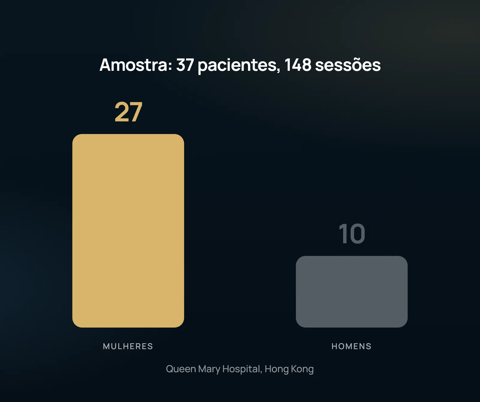
The control was taken seriously: to ensure that the plate on the uncooled side was at room temperature, it was heated with warm gauze and tested in the operator's forearm before the procedure. The cryogen (tetrafluoretane) was driven by piping to the handpiece, whose sapphire window had a circular opening of 4 cm in diameter crossed by the beam, with a viewing area of 2 cm and a real-time temperature monitor. The skin was cooled at 4 °C. All patients received EMLA under occlusion with tegaderm two hours before.
fluence was tested, not arbitrated
The dose tests started at 5 J/cm2, with increments of 0.5 J/cm2, immediately next to the marking. The test was interrupted when purpura or erosion appeared, and optimal fluence was defined as the highest fluence that caused immediate bleaching without these effects. There were two adjacent series — one with cooling on, the other with it off. Beam diameter of 2–3 mm and repeat rate of 3.3 Hz in both halves. The responses were collected by a trained nurse immediately after treatment and one week later, on a visual scale of 10 cm (0 to 10), with Student's t-test analysis.
What has changed and what hasn't changed
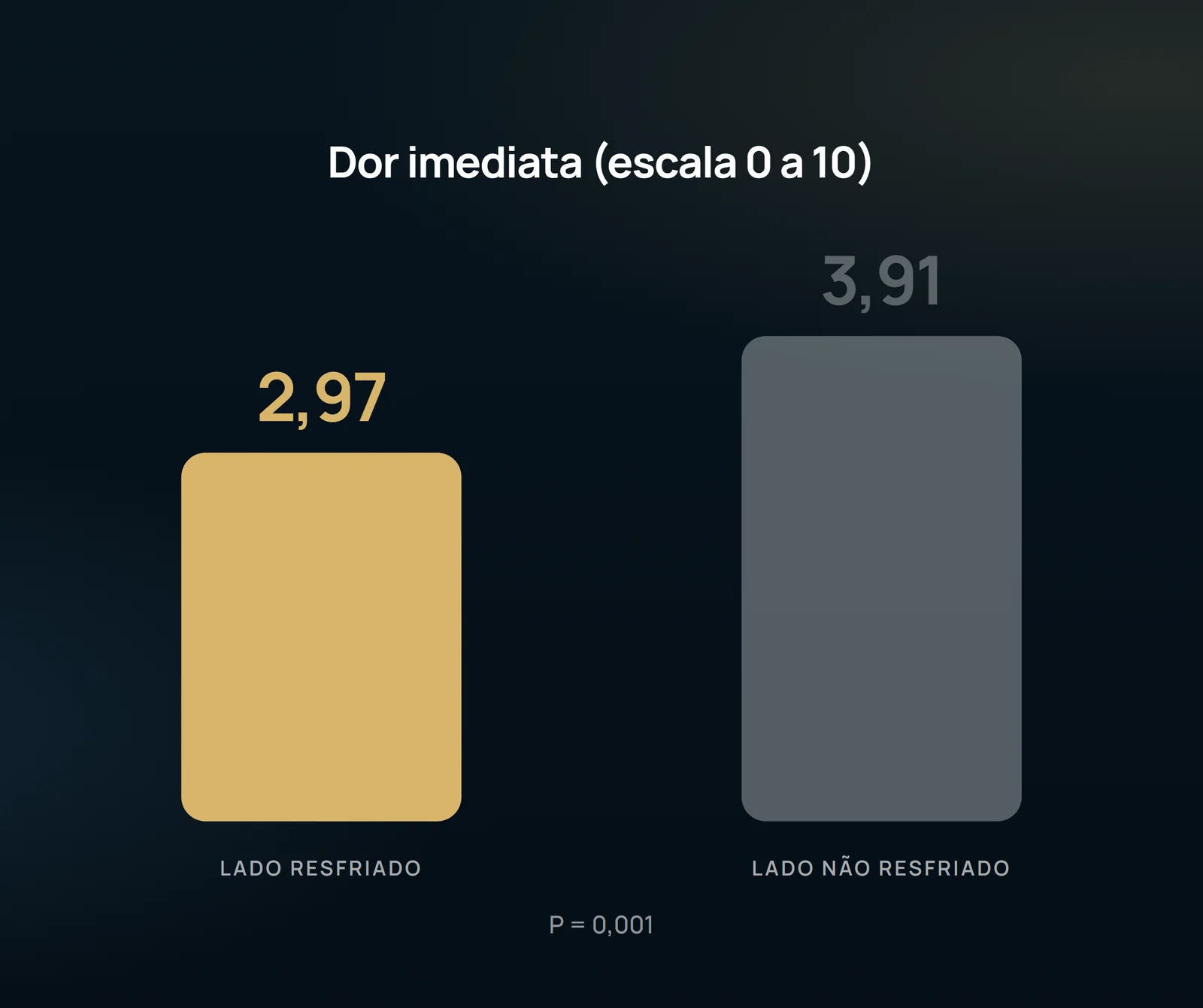
Eighty-two percent of the patients preferred the cold side; among the few who preferred the other side, the alleged motive was also less pain. However, there was no significant difference in fluence used, nor in discomfort, edema and pain one week after laser surgery.
Why the cold didn't buy power
The authors' explanation is physical. In a Q-switched system, the pulse duration is shorter than the heat relaxation time of the target: a temperature gradient is created between the chromophore and the neighboring tissue and, when this gradient collapses, localized shock waves are generated that fragment the target. This is the photomechanical effect, and cooling has no influence on it — unlike what happens with vascular lasers, where cooling acts on a thermal effect. In the vascular field, the article itself recalls the work of Chess and Chess, who treated 13 patients with lower limb telangiectasis with simultaneous cooling at firing and recorded a reduction in thermal injury to non-vascular tissue.
What the study does not authorize to state
The population is Ota nevus — dermal melanocytic lesion — and not venous disease: nothing here is evidence of lower limb telangiectasia, varicose veins or intravenous ablation, and direct extrapolation would be undue. There are 37 patients from a single hospital, with outcome collected by a questionnaire of pain, edema, discomfort, clearance and bleeding — there is no evaluation of scarring, late pigment change or long-term clearance. The negative conclusion is also specific to the system: the finding is that cooling did not allow greater fluence in a Q-switched laser, and the authors themselves point out that cryogen in spray with longer jet duration than that used for epidermal cooling would still require further studies. What you take to the office is a question, not a conduct: in the device you use, is the predominant effect thermal or photomechanical? That depends on what the cooler is actually buying.
Content addressed exclusively to health professionals. No individual result is guaranteed: the indication depends on clinical evaluation and imaging examination of each patient (CFM 2,336/2023).