Closed or merely thrombosed: the venous scaffold that allows a vein to reopen
Whiteley's review orders varicose veins by one criterion: whether or not the damage crosses the vein wall. When it does not cross, early occlusion is thrombus, and medium and adventitious survive as a living venous skeleton that allows recanalization.
 View the video lesson
The question that organizes Whiteley’s review is not what technique closes the vein — almost all of them close for a while. That's how far inside the wall the damage has come. It is with this ruler that the article orders intravenous thermal ablation, sclerotherapy with foam and open surgery, and it is the same rule that explains why foam, defeated in the trunk, is the best choice in other veins.
Target is transmural.
The biological outcome sought by intravenous thermal ablation is transmural death of the vein wall, with permanent fibrosis and atrophy of the treated segment. The article conditions this result to two factors mentioned together: the optimal amount of energy — measured as linear intravenous energy density — administered in optimal time. Energy and time, not just energy.

Inadequate treatment produces the opposite: damage restricted to the endothelium and to the intimate. The vein appears to be successful closure because thrombus is formed inside it — and it is exactly this picture that early control records as success.
The venous skeleton
What happens next is the core of the argument. The preserved medium and adventitia act as a living venous skeleton, and it is this skeleton that allows recanalization. The vein does not open by chance or because of equipment failure: it reopens because the structure that sustains it has never been reached.
Why foam is second line in trunk
Ultrasound-guided foam sclerotherapy is the second-line treatment for the incompetent stem vein. The justification presented is wall: both laboratory models of sclerotherapy in thick-walled veins and clinical studies showed lower long-term occlusion compared to intravenous thermal ablation. The foam is more successful when administered by a catheter, rather than injected from a single point, and with tumescence around the vein to increase the contact time of the sclerosing — but in the words of the article, this reduces the simplicity of the procedure and makes it similar to thermal ablation, with lower results.
And why, in other veins, foam is the ideal treatment
The same ruler reverses the result when the wall is thin. In small-diameter veins and thin walls — small varicosities and reticular veins, neovascular tissue, stripping tract revascularization and tissue of primary avalvular venous anomalies — foam sclerotherapy is, according to the article, the ideal treatment at the moment. Phlebectomy is inadequate in most of these veins, and they are too small and superficial for thermal ablation.
Open surgery and stripping
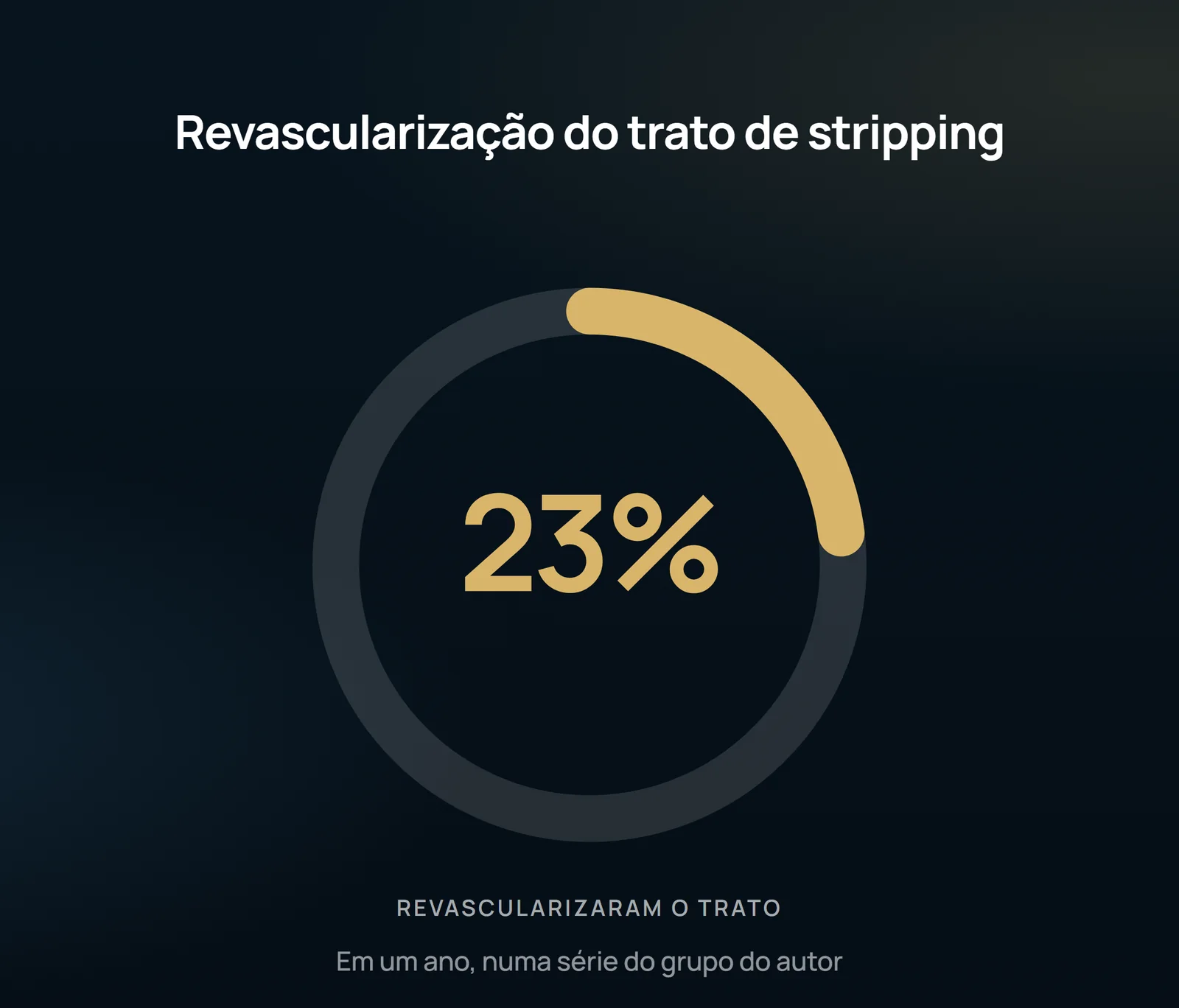
High stripping ligature is the recommended third line treatment after thermal ablation and foaming. In addition to the pain and discomfort of the procedure, the argument is new biological: the revascularization of the stripping tract. Surgical removal does not remove the vein definitively, because most of it grows again during healing — and returns without valves, reproducing the same recurrence problem.
Mechanochemical and cyanoacrylate: the same ruler
The two available mechanochemical ablation devices work by the principle of mechanical damage to the vein wall to increase the effect of sclerotherapy — i.e. by trying to ensure that chemical ablation becomes transmural. None of them are designed to treat piercing veins. The ablation with cyanoacrylate glue is rapid and does not require tumescence, and can be used directly by needle in a perforator; on the other hand, the main product is expensive against thermal ablation, and there are reports of inflammation at times very severe and extension to deep veins.
What the review does not allow to state
This is a narrative review of a unique author, who declares in the article himself to be founder of the College of Phlebology — it is not a systematic review with meta-analysis, and the selection of evidence is by the author. The text names the density of linear intravenous energy and the optimal time, but does not publish the value of either of the two: those who need the dose must go to the cited references. The venous skeleton is an explanation of mechanism, not an outcome measured in a randomized trial. The two percentages of revascularization of the stripping treatment come from series of the author's own group, in different follow-up, and not from an independent multicenter trial. And the article itself records that mechanochemical ablation and cyanoacrylate still need to prove cost-effectiveness in the face of established thermal techniques. None of this is a promise of outcome: the indication and choice of the technique depend on individual evaluation with duplex ultrasound, made by vascular surgeon.
Content addressed exclusively to health professionals. No individual result is guaranteed: the indication depends on clinical evaluation and imaging examination of each patient (CFM 2,336/2023).